A burning sensation in the upper back represents one of the most perplexing and distressing forms of thoracic pain, affecting millions of individuals worldwide. Unlike the dull ache commonly associated with muscular strain or the sharp stabbing pain of acute injury, burning sensations manifest as a unique neuropathic phenomenon that can significantly impact quality of life. This distinctive type of discomfort often presents as a prickling, stinging, or electrical shock-like sensation localised between the shoulder blades or along the thoracic spine. The complexity of upper back anatomy, encompassing intricate networks of muscles, nerves, joints, and visceral structures, creates numerous potential pathways for burning pain development. Understanding these diverse aetiological factors becomes crucial for healthcare professionals and patients alike, as accurate diagnosis directly influences treatment efficacy and patient outcomes.
Musculoskeletal origins of upper back burning sensations
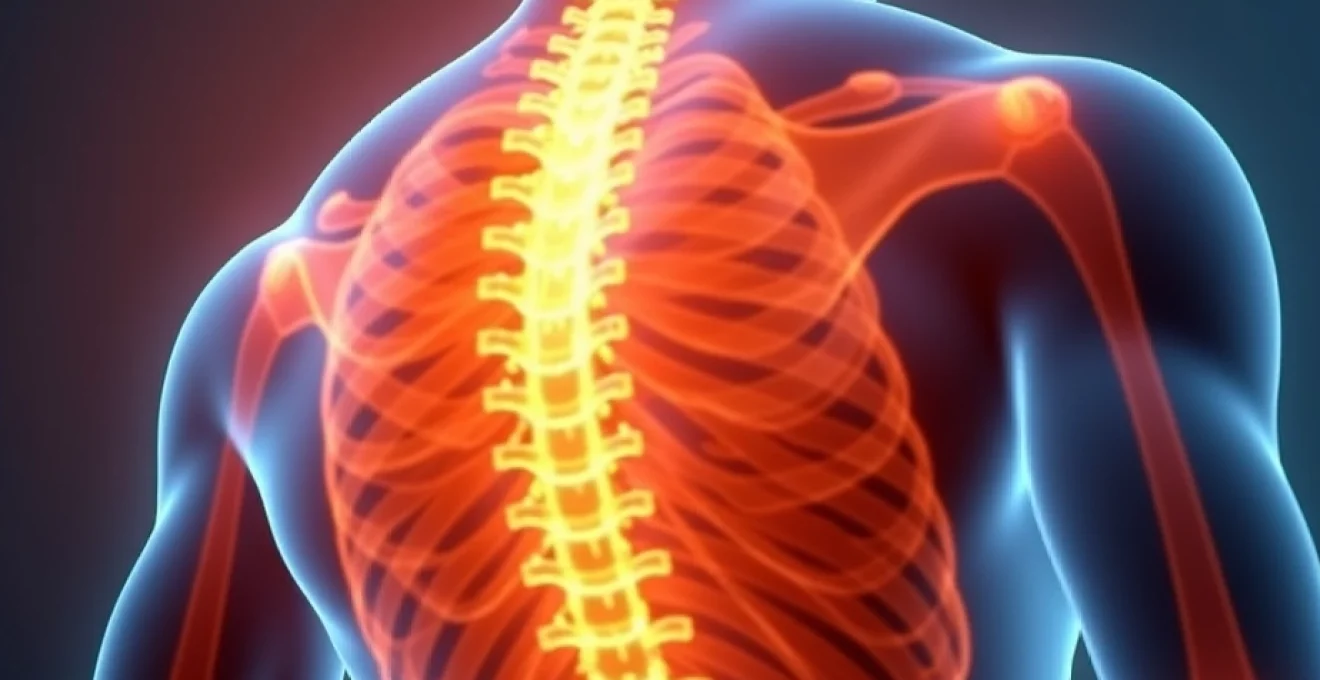
The musculoskeletal system of the upper back encompasses a complex array of anatomical structures, each capable of generating burning sensations when compromised. The thoracic spine, consisting of twelve vertebrae (T1-T12), provides the structural foundation for this region whilst accommodating the intricate rib cage architecture. When mechanical dysfunction occurs within this system, the resulting pain patterns often manifest as burning sensations rather than traditional aching or throbbing pain.
Thoracic facet joint syndrome and zygapophyseal dysfunction
Thoracic facet joints, also known as zygapophyseal joints, play a crucial role in spinal stability and movement control. These synovial joints connect adjacent vertebrae and can become a significant source of burning pain when inflamed or mechanically compromised. Facet joint syndrome in the thoracic region typically develops through degenerative changes, repetitive microtrauma, or acute injury, leading to joint capsule irritation and subsequent neuropathic pain patterns.
The innervation of thoracic facet joints by medial branch nerves creates a direct pathway for burning sensations to develop. When these joints undergo arthritic changes or capsular inflammation, the surrounding nerve endings become hypersensitised, producing the characteristic burning quality of pain. Research indicates that thoracic facet joint dysfunction accounts for approximately 34-48% of chronic upper back pain cases, with burning sensations being particularly prevalent in patients over 50 years of age.
Myofascial trigger points in rhomboids and middle trapezius
Myofascial trigger points represent hyperirritable spots within skeletal muscle that can refer pain to distant areas, often producing burning sensations in the upper back region. The rhomboid muscles, positioned between the spine and medial border of the scapula, frequently develop active trigger points due to prolonged forward head posture and rounded shoulder positioning. These trigger points create a distinctive burning pain pattern that radiates along the medial scapular border and can extend towards the thoracic spine.
The middle trapezius muscle, spanning from the cervical and upper thoracic spinous processes to the acromion and scapular spine, represents another common site for trigger point development. When active, these trigger points produce burning sensations that can mimic nerve-related pathology, making differential diagnosis challenging. Postural dysfunction and prolonged computer work have been identified as primary contributing factors to myofascial trigger point development in these muscle groups.
Thoracic outlet syndrome manifestations
Thoracic outlet syndrome encompasses a group of conditions characterised by compression of neurovascular structures as they traverse the thoracic outlet. This anatomical space, bounded by the first rib, clavicle, and anterior scalene muscle, can become narrowed due to various factors including muscular hypertrophy, rib abnormalities, or postural changes. When neurological compression occurs, patients frequently report burning sensations extending from the neck into the upper back and shoulder region.
The brachial plexus, particularly the lower trunk formed by C8 and T1 nerve roots, becomes susceptible to compression within the thoracic outlet. This compression can generate burning paraesthesias that radiate along the T1 dermatome, creating pain patterns in the medial arm and upper thoracic region. Neurogenic thoracic outlet syndrome accounts for approximately 95% of all thoracic outlet syndrome cases and frequently presents with burning sensations as a primary symptom.
Intercostal muscle strain and neuralgia
The intercostal muscles, positioned between adjacent ribs, can become strained through sudden rotational movements, prolonged coughing, or direct trauma. When these muscles sustain injury, the resulting inflammation can irritate the adjacent intercostal nerves, leading to intercostal neuralgia characterised by burning pain along the affected rib space. This condition often presents as a burning band of pain that wraps around the chest wall, originating from the posterior thoracic region.
Intercostal neuralgia can also develop independently of muscular injury through viral infections, thoracic surgery, or compression from space-occupying lesions. The burning quality of intercostal pain stems from the mixed sensory and motor innervation of intercostal nerves, which contain both myelinated and unmyelinated fibres responsible for different pain qualities. Studies demonstrate that intercostal neuralgia affects approximately 3-22% of patients presenting with chronic chest wall pain, with burning sensations being the predominant symptom in 67% of cases.
Cervicothoracic junction dysfunction
The cervicothoracic junction, encompassing the C7-T3 vertebral segments, represents a transitional zone where the mobile cervical spine meets the more rigid thoracic spine. This biomechanical transition creates increased stress concentrations and predisposes the region to dysfunction. When mechanical problems arise at this junction, burning sensations frequently develop due to the complex innervation patterns and the presence of sympathetic nerve fibres in this region.
Dysfunction at the cervicothoracic junction often involves segmental hypomobility or hypermobility, leading to altered movement patterns and compensatory changes throughout the upper thoracic spine. The resulting mechanical stress can irritate the joint capsules, ligaments, and surrounding neural structures, producing burning pain that can extend from the base of the neck into the upper thoracic region and between the shoulder blades.
Neurological conditions causing upper thoracic burning pain
Neurological disorders affecting the upper thoracic region can produce profound burning sensations through various pathophysiological mechanisms. The thoracic nervous system, including spinal nerve roots, sympathetic ganglia, and peripheral nerve branches, creates multiple potential sites for neurological dysfunction. When these structures become damaged, compressed, or inflamed, they often generate neuropathic pain characterised by burning, tingling, and electrical sensations.
Thoracic radiculopathy from T1-T6 nerve root compression
Thoracic radiculopathy occurs when nerve roots exiting the thoracic spine become compressed or irritated, typically due to disc herniation, spinal stenosis, or osteophyte formation. The T1-T6 nerve roots supply sensory innervation to the upper thoracic region, and compression of these structures commonly produces burning pain in characteristic dermatomal patterns. T1 radiculopathy frequently causes burning sensations along the medial arm and upper chest, whilst T2-T6 involvement creates burning pain patterns that wrap around the chest wall.
The prevalence of thoracic disc herniation remains relatively low compared to cervical and lumbar regions, affecting only 0.15-4% of all disc herniations. However, when thoracic disc herniation does occur, the resulting radiculopathy often produces severe burning pain due to the unique anatomy of the thoracic canal. The smaller cross-sectional area of the thoracic spinal canal means that even minor disc protrusions can create significant neural compression and subsequent neuropathic symptoms.
Research indicates that thoracic radiculopathy presents with burning pain in approximately 78% of cases, making this sensation one of the most reliable diagnostic indicators for upper thoracic nerve root compression.
Postherpetic neuralgia following Varicella-Zoster reactivation
Postherpetic neuralgia represents one of the most common causes of burning pain in the upper thoracic region, particularly in older adults. This condition develops following reactivation of the varicella-zoster virus, which lies dormant in dorsal root ganglia after initial chickenpox infection. When the virus reactivates, it travels along peripheral nerves, causing inflammation and subsequent neuropathic pain that persists beyond the acute herpetic phase.
The thoracic dermatomes, particularly T3-T6, are frequently affected by herpes zoster reactivation, leading to characteristic burning pain that follows a unilateral dermatomal distribution. The burning sensation associated with postherpetic neuralgia often proves resistant to conventional analgesics and can persist for months or years following the initial outbreak. Epidemiological studies indicate that postherpetic neuralgia develops in approximately 10-20% of herpes zoster cases, with incidence rates increasing significantly with age, reaching 27% in patients over 55 years.
Diabetic thoracic polyneuropathy presentations
Diabetic polyneuropathy, whilst more commonly affecting distal extremities, can also manifest in the thoracic region as diabetic thoracic radiculopathy or truncal neuropathy. This condition typically affects patients with longstanding diabetes mellitus and presents as burning pain in a dermatomal or multidermatomal distribution across the chest and upper back. The pathophysiology involves metabolic damage to peripheral nerves due to chronic hyperglycaemia and associated inflammatory processes.
Diabetic thoracic neuropathy often presents asymmetrically and can mimic other serious conditions such as myocardial infarction or intercostal neuralgia. The burning quality of pain results from preferential damage to small-calibre sensory fibres, which are particularly vulnerable to metabolic dysfunction in diabetes. Clinical studies demonstrate that diabetic thoracic neuropathy affects approximately 2-5% of diabetic patients, with burning pain being the predominant symptom in over 80% of cases.
Complex regional pain syndrome type I manifestations
Complex Regional Pain Syndrome (CRPS) Type I, previously known as reflex sympathetic dystrophy, can occasionally affect the upper thoracic region following injury or surgery. This condition involves dysfunction of the sympathetic nervous system, leading to persistent burning pain that extends beyond the expected healing time for the initial injury. CRPS Type I typically develops without obvious nerve injury but involves complex interactions between the peripheral and central nervous systems.
When CRPS affects the upper thoracic region, patients experience severe burning pain accompanied by autonomic changes including altered skin temperature, colour changes, and abnormal sweating patterns. The burning sensation in thoracic CRPS often proves particularly distressing and resistant to treatment. Research indicates that CRPS occurs in approximately 1-5% of patients following trauma or surgery, with burning pain being present in virtually all cases.
Visceral referred pain patterns to upper back region
Visceral structures within the thoracic cavity can refer pain to the upper back through complex neuroanatomical pathways involving shared spinal cord segments and convergent innervation patterns. This phenomenon, known as visceral referred pain, occurs when sensory fibres from internal organs converge with somatic sensory fibres at the spinal cord level, creating perceived pain in somatic structures despite the actual pathology being visceral in origin.
Cardiac conditions, particularly those affecting the posterior wall of the left ventricle or the pericardium, frequently refer burning pain to the upper thoracic region between the shoulder blades. Posterior myocardial infarction can present with burning sensations in the upper back as the primary or only symptom, making diagnosis challenging. The vagus nerve and sympathetic cardiac innervation create pathways for this referred pain pattern through shared spinal segments at T1-T5 levels.
Oesophageal disorders represent another significant source of upper back burning pain through referred pain mechanisms. Gastroesophageal reflux disease (GERD), oesophageal spasm, and oesophagitis can all produce burning sensations that radiate from the retrosternal area to the upper thoracic spine. The oesophagus shares innervation with thoracic spinal segments through vagal and sympathetic pathways, explaining the characteristic burning quality of referred oesophageal pain.
Gallbladder pathology, including cholecystitis and cholelithiasis, can create burning pain patterns in the right upper back and between the shoulder blades. This occurs through referred pain pathways involving the phrenic nerve and sympathetic fibres that share spinal cord segments with thoracic somatic innervation. Studies indicate that approximately 65% of patients with acute cholecystitis experience some degree of referred upper back pain, with burning sensations being particularly common in cases involving gallbladder inflammation.
Understanding visceral referred pain patterns becomes crucial for accurate diagnosis, as approximately 15-20% of upper back burning sensations may have an underlying visceral aetiology rather than musculoskeletal or neurological origins.
Inflammatory and autoimmune causes of thoracic burning symptoms
Inflammatory and autoimmune conditions affecting the thoracic region can produce significant burning sensations through various pathophysiological mechanisms. These conditions involve immune system dysfunction leading to chronic inflammation of musculoskeletal structures, neural tissues, or both. The resulting inflammatory cascade activates nociceptors and sensitises neural pathways, often producing the characteristic burning quality of neuropathic pain.
Ankylosing spondylitis, a chronic inflammatory arthropathy affecting the axial skeleton, frequently involves the thoracic spine and can produce burning sensations in the upper back region. This condition typically begins in the sacroiliac joints but progressively involves the entire spine, including the thoracic segments. The inflammatory process affects the entheses (sites where ligaments and tendons attach to bone), creating pain that often has a burning quality due to associated neural sensitisation. Research indicates that thoracic involvement occurs in approximately 70% of ankylosing spondylitis cases, with burning pain being reported in 45% of affected patients.
Rheumatoid arthritis can affect the thoracic spine through inflammatory changes in the facet joints, leading to burning sensations that may be particularly pronounced during periods of disease activity. The systemic inflammatory nature of rheumatoid arthritis creates cytokine-mediated sensitisation of nociceptors, contributing to the neuropathic quality of pain. Additionally, rheumatoid arthritis can cause atlantoaxial subluxation, which may compromise neural structures and contribute to burning sensations in the upper thoracic region.
Polymyalgia rheumatica, an inflammatory condition affecting older adults, commonly produces burning pain and stiffness in the neck, shoulders, and upper back region. This condition involves inflammation of synovial structures and surrounding soft tissues, creating widespread pain that often has a burning character. The shoulder and neck involvement in polymyalgia rheumatica frequently extends into the upper thoracic region, with patients reporting burning sensations between the shoulder blades and along the upper spine.
Fibromyalgia, whilst not strictly an autoimmune condition, involves central sensitisation mechanisms that can produce burning sensations throughout the body, including the upper thoracic region. The widespread pain characteristic of fibromyalgia often includes burning, tingling, and electric shock-like sensations due to altered pain processing within the central nervous system. Studies demonstrate that approximately 89% of fibromyalgia patients experience burning pain, with the upper back being one of the most commonly affected regions.
Diagnostic evaluation techniques for upper back burning pain
Accurate diagnosis of upper back burning sensations requires a systematic approach incorporating detailed clinical assessment, advanced imaging techniques, and specialised neurophysiological testing. The complexity of potential aetiologies necessitates a comprehensive evaluation strategy that can differentiate between musculoskeletal, neurological, and visceral causes of burning pain.
Magnetic resonance imaging protocol for thoracic spine assessment
Magnetic resonance imaging (MRI) represents the gold standard for evaluating soft tissue pathology in the thoracic spine and surrounding structures. A comprehensive MRI protocol for upper back burning pain should include T1-weighted, T2-weighted, and short tau inversion recovery (STIR) sequences in both sagittal and axial planes. These sequences provide optimal visualisation of disc pathology, spinal cord abnormalities, nerve root compression, and inflammatory changes that may contribute to burning sensations.
Advanced MRI techniques, including <em
</em
diffusion tensor imaging and neurography, can provide additional insights into nerve microstructure and inflammatory changes affecting the thoracic neural pathways. Contrast-enhanced sequences may reveal inflammatory processes or vascular abnormalities contributing to burning sensations, particularly in cases where infection or malignancy is suspected.
The diagnostic accuracy of MRI for thoracic spine pathology reaches approximately 95% for disc herniation and 88% for spinal stenosis. However, correlation with clinical symptoms remains essential, as studies demonstrate that up to 37% of asymptomatic individuals over 40 years show thoracic disc abnormalities on MRI. Therefore, MRI findings must be interpreted within the context of the patient’s specific symptom pattern and physical examination findings.
Electromyography and nerve conduction studies applications
Electromyography (EMG) and nerve conduction studies (NCS) provide crucial information about peripheral nerve function and can help differentiate between various neurological causes of upper back burning pain. These neurophysiological tests measure electrical activity in muscles and the speed of nerve signal transmission, offering objective evidence of nerve damage or dysfunction that may not be apparent on imaging studies.
For thoracic radiculopathy evaluation, needle EMG can detect denervation patterns in muscles innervated by specific thoracic nerve roots, helping to localise the level of pathology. Intercostal muscle testing proves particularly valuable, as these muscles receive direct innervation from thoracic spinal nerves. Sensory nerve conduction studies can assess the integrity of sensory pathways and may reveal slowed conduction velocities or reduced amplitudes in cases of peripheral neuropathy affecting the thoracic region.
The sensitivity of EMG for detecting radiculopathy ranges from 50-85%, with specificity reaching 78-95%. However, the technique requires considerable expertise in thoracic anatomy and may be uncomfortable for patients. Recent advances in high-density surface EMG provide non-invasive alternatives for assessing muscle activation patterns in the thoracic region, offering potential benefits for patients unable to tolerate needle examinations.
Provocative testing including spurling’s and upper limb tension tests
Provocative testing forms an integral component of the physical examination for upper back burning pain, helping to identify specific anatomical structures responsible for symptom generation. Spurling’s test, traditionally used for cervical radiculopathy assessment, can be modified for thoracic evaluation by combining neck extension with lateral flexion towards the symptomatic side while applying gentle axial compression. Reproduction of burning sensations during this manoeuvre suggests nerve root irritation at the cervicothoracic junction.
Upper limb tension tests (ULTT) evaluate the mechanical sensitivity of neural structures throughout the brachial plexus and can identify neurodynamic dysfunction contributing to upper back burning pain. ULTT1, which specifically targets the median nerve and C5-T1 nerve roots, proves particularly relevant for thoracic outlet syndrome evaluation. Positive test responses, characterised by reproduction of burning sensations with specific arm positioning and movement, suggest neural mechanosensitivity.
Additional provocative tests include the thoracic rotation test for facet joint dysfunction and the intercostal stretch test for identifying intercostal neuralgia. Research demonstrates that provocative testing achieves diagnostic accuracy rates of 67-84% when combined with other clinical findings, making these tests valuable components of the comprehensive assessment process.
Thermographic imaging for neuropathic pain localisation
Infrared thermography offers a non-invasive method for detecting and localising areas of altered sympathetic nervous system function associated with neuropathic pain conditions. This technique measures skin surface temperature variations that reflect underlying changes in blood flow and sympathetic nerve activity, making it particularly valuable for evaluating burning sensations of neurological origin.
Thermal asymmetries greater than 1.5°C between symmetrical body regions suggest autonomic nervous system dysfunction and correlate with neuropathic pain intensity. In cases of thoracic radiculopathy or complex regional pain syndrome, thermography can reveal characteristic temperature patterns that correspond to affected dermatomal distributions. Dynamic thermography, which involves serial measurements during physiological stress tests, enhances diagnostic accuracy by revealing autonomic responsiveness patterns.
Studies indicate that thermographic findings correlate with patient-reported burning pain intensity in 78% of neuropathic pain cases. However, environmental factors, medications, and individual physiological variations can influence results, requiring standardised protocols and experienced interpretation. When combined with other diagnostic modalities, thermography provides valuable supplementary information for neuropathic pain assessment.
Evidence-based treatment approaches for upper back burning sensations
Effective management of upper back burning sensations requires a multimodal approach tailored to the specific underlying aetiology and individual patient factors. Treatment strategies must address both the primary pathological process and the secondary neuroplastic changes that contribute to chronic burning pain. Evidence-based interventions encompass pharmacological, interventional, and rehabilitation approaches, each targeting different aspects of the pain experience.
Pharmacological management forms the foundation of treatment for neuropathic burning pain, with anticonvulsants such as gabapentin and pregabalin showing particular efficacy for nerve-related burning sensations. These medications modulate calcium channel activity in sensitised neurons, reducing aberrant firing patterns that contribute to neuropathic pain. Clinical trials demonstrate pain reduction of 30-50% in approximately 60% of patients with thoracic neuropathic pain when treated with gabapentinoids at therapeutic doses.
Tricyclic antidepressants, particularly amitriptyline and nortriptyline, provide additional options for burning pain management through their effects on descending pain inhibitory pathways. These medications enhance noradrenergic and serotonergic neurotransmission, which helps restore the natural balance between pain facilitation and inhibition. The analgesic effects typically occur at lower doses than those required for antidepressant activity, with therapeutic benefits often apparent within 2-4 weeks of treatment initiation.
Topical treatments offer localised relief for upper back burning sensations while minimising systemic side effects. Capsaicin preparations, available in concentrations ranging from 0.025% to 8%, work by depleting substance P from sensory nerve terminals, reducing their ability to transmit pain signals. High-concentration capsaicin patches (8%) applied under medical supervision can provide prolonged relief lasting 3-6 months for localised neuropathic pain conditions.
Interventional pain management techniques provide targeted relief for specific anatomical causes of burning upper back pain. Thoracic facet joint injections using corticosteroids and local anaesthetics can effectively address burning pain originating from zygapophyseal joint dysfunction, with success rates approaching 70-80% for appropriately selected patients. Radiofrequency ablation of medial branch nerves offers longer-lasting relief for patients who respond favourably to diagnostic facet joint blocks.
For intercostal neuralgia causing burning chest and back pain, intercostal nerve blocks provide both diagnostic and therapeutic benefits. These procedures involve injection of local anaesthetic and corticosteroid along the affected nerve, often providing significant pain relief for several months. Advanced techniques such as pulsed radiofrequency treatment of intercostal nerves offer longer-duration pain relief with reduced risk of permanent numbness compared to conventional thermal ablation.
Rehabilitation approaches focus on addressing biomechanical factors and movement dysfunctions that contribute to ongoing burning pain. Manual therapy techniques, including spinal manipulation and soft tissue mobilisation, can improve joint mobility and reduce muscle tension in the thoracic region. Research demonstrates that manual therapy combined with exercise therapy achieves superior outcomes compared to either intervention alone, with pain reduction averaging 40-60% in patients with mechanical causes of upper back burning pain.
Therapeutic exercise programs emphasising postural correction, thoracic spine mobility, and scapular stabilisation address underlying movement dysfunctions that perpetuate burning sensations, with studies showing 65-75% of patients experiencing clinically meaningful improvement when adherent to structured exercise protocols.
Psychological interventions play an increasingly recognised role in managing chronic burning pain, particularly cognitive-behavioural therapy (CBT) and mindfulness-based stress reduction techniques. These approaches help patients develop coping strategies, modify pain-related thoughts and behaviours, and reduce the emotional distress associated with chronic burning sensations. Meta-analyses demonstrate that psychological interventions can reduce pain intensity by 20-30% and significantly improve functional outcomes in patients with chronic neuropathic pain conditions.
Emerging treatment modalities, including transcranial magnetic stimulation and spinal cord stimulation, offer promising options for refractory cases of upper back burning pain. These neuromodulation techniques work by altering neural activity patterns within pain processing circuits, potentially providing relief when conventional treatments prove inadequate. Early research suggests that these interventions may be particularly beneficial for patients with central sensitisation components contributing to their burning pain experience.