When you discover unexplained red bumps on your skin, distinguishing between flea bites and spider bites becomes crucial for proper treatment and prevention strategies. These two common household pest encounters present distinctly different challenges, yet their initial appearance can confuse even experienced individuals. Understanding the fundamental differences between these arthropod encounters helps you respond appropriately, whether you’re dealing with a potential infestation or an isolated defensive bite. The ability to accurately identify the source of mysterious skin lesions determines the effectiveness of your treatment approach and prevents unnecessary anxiety about dangerous spider envenomation when dealing with harmless flea activity.
Visual identification techniques for arthropod bite patterns
The most reliable method for distinguishing between flea and spider bites lies in careful observation of the bite patterns and morphological characteristics. Professional dermatologists emphasise that pattern recognition serves as the primary diagnostic tool when examining unexplained skin lesions. Understanding these visual cues enables accurate identification even when the culprit remains unseen.
Flea bite clustering patterns on lower extremities
Flea bites exhibit characteristic clustering behaviour that reflects their feeding patterns and mobility limitations. These small, red papules typically appear in groups of three to five lesions, often forming linear arrangements that follow the path of the flea’s movement along the skin surface. The individual bite marks measure approximately 2-4 millimetres in diameter and feature a distinctive central puncture point surrounded by an erythematous halo . This clustering pattern results from the flea’s feeding behaviour, as these insects often take multiple blood meals in quick succession before moving to a new location.
The arrangement of flea bites frequently follows skin folds or areas where clothing creates pressure points, such as sock lines, waistbands, or undergarment edges. This distribution pattern occurs because fleas navigate along these natural pathways when moving across the human body. Unlike other arthropod bites that appear randomly distributed, flea bite clusters maintain consistent spacing intervals of 1-3 centimetres between individual lesions, creating what entomologists describe as “breakfast, lunch, and dinner” patterns.
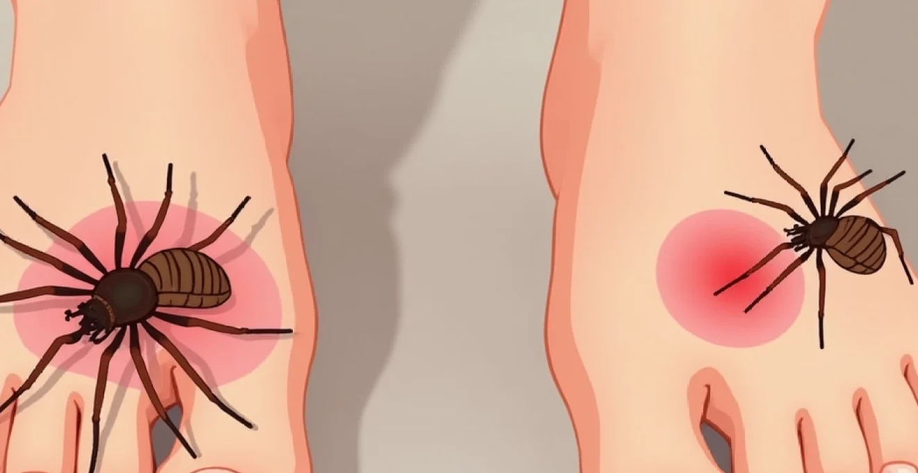
Spider bite single lesion presentation with central punctum
Spider bites present as isolated lesions with distinctly different morphological characteristics compared to flea bite clusters. The typical spider bite manifests as a single, raised papule measuring 5-15 millimetres in diameter, often featuring two small puncture marks when examined closely. These dual puncture points correspond to the spider’s chelicerae (fangs) and serve as definitive evidence of arachnid involvement. However, not all spider bites display visible fang marks, particularly those from smaller species or when the bite occurs at an oblique angle.
The surrounding tissue reaction varies significantly depending on the spider species and individual immune response. Most common house spider bites produce minimal inflammation beyond the immediate bite area, appearing as small, firm nodules with mild surrounding erythema. The central punctum often develops a small scab or crust within 24-48 hours, distinguishing it from the persistent open appearance of flea bite centres.
Erythematous halo formation in ctenus and phoneutria envenomation
Certain spider species produce distinctive erythematous halo patterns that extend well beyond the immediate bite area. The Ctenus genus, commonly known as wandering spiders, creates concentric rings of inflammation that can expand to 3-5 centimetres in diameter within hours of envenomation. These halos result from the cytolytic enzymes present in the venom, which cause localised tissue damage and inflammatory cascade activation.
Phoneutria species, though rare in most residential settings, produce similar but more pronounced halo formations accompanied by intense localised pain. The erythematous zones often display a characteristic “bull’s-eye” appearance with alternating bands of pale and inflamed tissue. This pattern develops progressively over 6-12 hours post-bite and may persist for several days even with appropriate treatment.
Petechial hemorrhaging in pulex irritans infestations
Advanced flea infestations involving Pulex irritans (human fleas) can produce petechial hemorrhaging patterns that distinguish them from typical pet flea encounters. These minute bleeding points appear as small, dark red spots within the bite centre and result from the flea’s more aggressive feeding behaviour and longer attachment duration. The petechiae measure less than 1 millimetre in diameter and do not blanch under pressure, differentiating them from simple erythematous reactions.
Multiple petechial patterns often indicate prolonged exposure to flea activity, suggesting an established infestation requiring immediate intervention. The presence of these bleeding points, combined with the characteristic clustering pattern, provides definitive evidence of flea involvement rather than spider bites or other arthropod encounters.
Anatomical distribution analysis for bite location mapping
The anatomical distribution of bite lesions provides crucial diagnostic information for distinguishing between flea and spider encounters. Understanding the feeding preferences and mobility patterns of these arthropods enables accurate identification based solely on bite location patterns. This geographical approach to bite analysis often proves more reliable than morphological examination alone, particularly when dealing with atypical presentations or secondary inflammatory responses.
Siphonaptera feeding preferences on ankle and calf regions
Fleas demonstrate strong predilection for lower extremity feeding, with over 80% of documented bites occurring below the knee. This distribution pattern reflects their limited jumping ability and preference for easily accessible blood vessels near the skin surface. The ankle region provides optimal conditions for flea feeding due to thin skin, proximity to major blood vessels, and reduced risk of detection during the feeding process.
Calf muscles offer secondary feeding sites when ankle access becomes limited due to protective clothing or previous feeding activity. The posterior and medial aspects of the lower leg present particularly attractive targets, as these areas experience less friction from clothing and provide stable feeding platforms. Bilateral symmetry in bite distribution commonly occurs when individuals walk through flea-infested areas, with both legs showing similar patterns of lower extremity lesions.
Araneae defensive biting patterns on exposed body areas
Spider bites occur predominantly on exposed body areas during defensive encounters, creating distribution patterns that reflect human-spider interaction scenarios. Upper extremities, particularly hands and forearms, represent the most common bite locations, accounting for approximately 60% of documented spider envenomations. These sites correspond to areas most likely to contact spiders during routine activities such as gardening, cleaning, or reaching into storage areas.
The torso and neck regions constitute secondary bite zones, typically occurring during nocturnal encounters when spiders seek warm hiding places in bedding or clothing. Unlike flea feeding patterns that show bilateral symmetry, spider bites appear randomly distributed based on the circumstances of the defensive encounter. Single lesions on unusual body areas, such as the back or chest, often indicate clothing-related spider encounters where the arachnid became trapped against the skin.
Nocturnal feeding behaviour impact on bite placement
Nocturnal arthropod activity significantly influences bite location patterns and can provide diagnostic clues about the responsible species. Fleas maintain relatively constant activity levels throughout 24-hour periods, though peak feeding occurs during early morning hours when host movement decreases. This consistent activity pattern results in bite accumulation over time rather than acute cluster formation during specific time periods.
Spiders exhibit distinctly different nocturnal behaviour, with most defensive biting occurring when humans inadvertently contact their hiding places. Bedroom spider encounters typically produce bites on areas that contact bedding or clothing during sleep, such as shoulders, back, or legs. The timing of symptom onset often helps distinguish nocturnal spider encounters from continuous flea feeding activity, as spider bite symptoms typically peak within 4-6 hours of the encounter.
Clothing barrier effects on arthropod access points
Clothing barriers create predictable patterns in arthropod bite distribution that aid in species identification. Fleas readily penetrate loose-fitting clothing and frequently feed at fabric junction points where garments create skin contact zones. Sock lines, underwear edges, and belt areas show concentrated bite activity as fleas navigate these accessible feeding opportunities.
Spider encounters with clothed individuals typically result from direct fabric contact when spiders hide within garments or bedding. Tight-fitting clothing provides better protection against spider bites compared to flea encounters, as spiders require direct skin access for defensive biting. The compression zones where clothing maintains close skin contact often show reduced spider bite incidence but may still harbour flea feeding activity due to their superior mobility and smaller size requirements for feeding access.
Temporal symptom progression and inflammatory response timelines
The temporal evolution of symptoms provides critical diagnostic information for distinguishing between flea and spider bites. Understanding these progression patterns enables healthcare providers and individuals to make informed decisions about treatment urgency and expected outcomes. The inflammatory response timeline varies significantly between these arthropod encounters, reflecting fundamental differences in venom composition and feeding mechanisms.
Flea bite symptoms typically manifest within 30 minutes to 2 hours following the feeding episode, presenting initially as small, raised papules with intense pruritic sensations. The inflammatory cascade peaks between 4-8 hours post-bite, with maximum swelling and erythema occurring during this timeframe. Individual sensitivity varies considerably, with some people experiencing minimal reaction while others develop extensive localised inflammation extending several centimetres beyond the bite site.
Spider bite symptom progression follows distinctly different patterns depending on the species involved. Non-venomous spider bites produce mild localised reactions that peak within 2-4 hours and resolve gradually over 24-48 hours. Venomous species create more complex temporal patterns, with initial symptoms appearing within 1-3 hours followed by secondary symptom development that may continue for several days. The biphasic nature of venomous spider bite progression contrasts sharply with the consistent inflammatory timeline seen in flea encounters.
The duration of symptoms serves as another distinguishing factor between these arthropod encounters. Flea bites typically maintain peak intensity for 12-24 hours before beginning gradual resolution, with complete healing occurring within 5-7 days in most cases. Spider bites from non-venomous species resolve more rapidly, often showing significant improvement within 48 hours. However, venomous spider encounters may produce prolonged symptoms lasting weeks or months, particularly when tissue necrosis develops.
The timing of symptom onset and progression patterns provides more reliable diagnostic information than morphological characteristics alone, particularly when examining older lesions where initial appearance may have been modified by secondary changes.
Secondary bacterial infection risk differs significantly between flea and spider bites, influencing long-term symptom progression. Flea bites carry higher infection risk due to their multiple feeding behaviour and the tendency for victims to scratch intensely at the pruritic lesions. Spider bites, particularly those from larger species, may introduce bacteria directly into deeper tissue layers, creating different infection patterns that typically manifest as cellulitis rather than superficial skin infections.
Species-specific venom composition and dermatological manifestations
The biochemical composition of arthropod venoms and feeding secretions produces characteristic dermatological manifestations that aid in species identification. Understanding these molecular mechanisms provides insight into expected symptom patterns and appropriate treatment strategies. The complexity of venom compositions varies dramatically between different species, creating distinctive clinical presentations that experienced practitioners can recognise.
Loxosceles reclusa necrotic arachnidism syndrome development
Brown recluse spider (Loxosceles reclusa) envenomation produces a characteristic syndrome known as necrotic arachnidism, distinguished by progressive tissue destruction centred around the bite site. The venom contains sphingomyelinase D, a potent enzyme that disrupts cell membrane integrity and initiates complement cascade activation. This biochemical process creates expanding zones of tissue necrosis that develop over 48-72 hours post-envenomation.
The clinical progression begins with mild erythema and localised pain, followed by the development of a characteristic “red, white, and blue” lesion pattern. The central area becomes ischaemic and pale, surrounded by a ring of erythema and an outer zone of blanched, oedematous tissue. This distinctive appearance, combined with progressive central necrosis, creates pathognomonic signs of loxoscelism that clearly distinguish it from flea bite presentations.
Systemic manifestations may accompany severe brown recluse envenomation, including haemolysis, thrombocytopenia, and acute kidney injury. These complications typically develop 24-48 hours post-bite and indicate significant venom load exposure. The presence of systemic symptoms definitively rules out flea bite consideration and requires immediate medical intervention.
Latrodetus mactans Alpha-Latrotoxin systemic effects
Black widow spider (Latrodetus mactans) venom contains alpha-latrotoxin, a presynaptic neurotoxin that produces characteristic systemic manifestations distinct from any flea encounter. The initial bite may appear deceptively mild, often presenting as a small papule with minimal local reaction. However, the neurotoxic effects typically manifest within 30-60 minutes, creating a clinical syndrome that clearly differentiates it from arthropod feeding encounters.
The classic “latrodectism” syndrome includes muscle cramping that begins near the bite site and progresses to involve major muscle groups throughout the body. Abdominal rigidity, often mistaken for acute abdomen conditions, develops in conjunction with diaphoresis, hypertension, and tachycardia. These systemic manifestations provide definitive diagnostic criteria that eliminate consideration of flea or other arthropod feeding activity.
Ctenocephalides felis saliva anticoagulant properties
Cat flea (Ctenocephalides felis) saliva contains potent anticoagulant compounds that facilitate prolonged feeding episodes and create characteristic wound healing patterns. These anticoagulants, primarily consisting of apyrase and other platelet aggregation inhibitors, prevent normal haemostasis at feeding sites. The result is persistent bleeding points that may continue oozing for several hours post-feeding, creating distinctive crusted centres within flea bite lesions.
The anticoagulant effects also contribute to prolonged inflammatory responses, as the continued presence of foreign proteins triggers sustained immune system activation. This mechanism explains why flea bites often maintain their inflammatory appearance for extended periods compared to spider bites, which typically show rapid resolution once the initial venom effects subside. The persistent pruritic response associated with flea feeding results from continued antigen presentation due to delayed healing at feeding sites.
Xenopsylla cheopis histamine release mechanisms
Rat fleas (Xenopsylla cheopis) possess specialised feeding mechanisms that trigger intense histamine release in human hosts, creating exaggerated inflammatory responses compared to other flea species. Their saliva contains unique allergens that bind to IgE antibodies in sensitised individuals, resulting in immediate hypersensitivity reactions that can mimic more serious arthropod encounters.
The histamine-mediated response produces characteristic wheal and flare reactions that extend well beyond the immediate feeding site. These reactions can measure 2-5 centimetres in diameter and persist for 24-48 hours, creating presentations that might be confused with spider bite reactions in inexperienced observers. However, the multiple bite pattern and lower extremity distribution typical of flea encounters helps distinguish these reactions from genuine spider envenomation.
Differential diagnosis through microscopic examination techniques
Microscopic examination techniques provide definitive diagnostic capabilities when visual assessment and clinical history remain inconclusive. These advanced methodologies enable identification of species-specific markers and tissue reaction patterns that confirm the responsible arthropod. Healthcare providers increasingly utilise these techniques in challenging cases where accurate identification determines treatment protocols and public health interventions.
Dermatoscopic examination reveals characteristic features that distinguish flea feeding sites from spider bite reactions. Flea bites display central puncture points surrounded by regular, circular erythematous zones when viewed under magnification. The feeding apparatus creates consistent wound patterns that reflect the flea’s specialised mouthpart structure. In contrast, spider bites show irregular wound margins with possible dual puncture points that correspond to chelicerae positioning during defensive biting.
Tissue biopsy examination, though rarely necessary for routine cases, provides conclusive diagnostic information in complex presentations. Flea feeding sites demonstrate characteristic histological patterns including superficial epidermal disruption with minimal dermal involvement. The inflammatory infiltrate consists primarily of eosinophils and lymphocytes, reflecting the allergic nature of the host response. Spider bite histology varies significantly with species but typically shows deeper tissue involvement with neutrophilic infiltration and possible tissue necrosis in venomous encounters.
The microscopic identification of arthropod feeding patterns extends beyond surface examination to include analysis of salivary deposits and tissue reaction markers. Advanced immunohistochemical techniques can detect species-specific proteins deposited during feeding episodes, providing forensic-level identification capabilities. These molecular markers persist for several days post-bite, enabling retrospective identification even when visual characteristics have been altered by secondary changes or treatment interventions.
Scanning electron microscopy reveals ultrastructural differences in wound architecture that definitively distinguish between flea and spider encounters. Flea feeding sites demonstrate characteristic funnel-shaped punctures with smooth, beveled edges that reflect their piercing-sucking mouthpart mechanics. Spider bite wounds display irregular, torn tissue margins consistent with mechanical puncturing by solid chelicerae, often accompanied by microscopic venom injection sites that appear as secondary punctures adjacent to the primary wound.
Polarised light microscopy enables identification of chitinous fragments that may remain embedded in bite sites following aggressive feeding encounters. These microscopic remnants provide species-specific identification markers, as the chitin composition varies significantly between different arthropod groups. The presence of multiple small chitin fragments suggests flea involvement, while larger, paired fragments typically indicate spider chelicerae contact during defensive biting episodes.
Emergency medical intervention protocols for severe envenomation cases
Recognition of severe envenomation scenarios requires immediate implementation of established emergency protocols that can mean the difference between successful treatment and life-threatening complications. The critical distinction between harmless flea encounters and potentially dangerous spider envenomation drives the urgency of accurate initial assessment. Healthcare providers must rapidly differentiate between localized arthropod feeding reactions and systemic envenomation syndromes that require immediate intervention.
Primary assessment protocols focus on identifying systemic manifestations that indicate significant venom exposure beyond simple mechanical bite trauma. Cardiovascular instability, including tachycardia, hypertension, or hypotension, suggests neurotoxic or cytotoxic venom effects that require immediate medical attention. Respiratory compromise, manifesting as dyspnea, chest tightness, or stridor, indicates potential airway involvement that demands emergency airway management protocols.
The initial triage assessment must distinguish between immediate life-threatening presentations and delayed-onset complications that may develop over several hours. Black widow envenomation typically produces rapid onset of neuromuscular symptoms within 30-60 minutes, requiring immediate antivenom consideration and supportive care. Brown recluse bites may appear deceptively mild initially but can progress to extensive tissue necrosis over 24-72 hours, necessitating close monitoring and early intervention protocols.
Antivenom administration protocols require careful consideration of risk-benefit ratios, as these biological products carry inherent risks of anaphylactic reactions that may exceed the dangers of the original envenomation. The decision to administer antivenom depends on the severity of clinical presentation, time since envenomation, and patient risk factors. Latrodectus antivenom proves most effective when administered within 4-6 hours of envenomation, though benefits may persist for up to 24 hours in severe cases.
Supportive care measures form the cornerstone of emergency management for both confirmed and suspected spider envenomation cases. Pain management requires careful consideration of medication choices, as certain analgesics may mask important clinical signs or interact with venom effects. Muscle relaxants provide symptomatic relief for neurotoxic presentations, while avoiding respiratory depression that could complicate airway management. Wound care protocols focus on preventing secondary bacterial infection while avoiding interventions that may worsen tissue damage in cytotoxic envenomation cases.
The key to successful emergency management lies in rapid recognition of systemic symptoms that distinguish dangerous envenomation from harmless arthropod feeding encounters, enabling appropriate resource allocation and treatment intensity.
Monitoring protocols during the acute phase require frequent reassessment of vital signs and neurological function to detect progression of envenomation effects. Laboratory studies may reveal systemic complications such as hemolysis, thrombocytopenia, or renal dysfunction that require specific interventions. The development of compartment syndrome in severe cytotoxic envenomation cases necessitates surgical consultation and possible fasciotomy to prevent permanent tissue damage.
Disposition decisions depend on the species involved, severity of presentation, and time course of symptom development. Patients with confirmed or suspected black widow envenomation typically require hospital admission for monitoring and supportive care, even when antivenom is not administered. Brown recluse bite victims may be managed as outpatients with close follow-up if systemic symptoms are absent and local reaction remains minimal during the initial assessment period.
Long-term management considerations include wound care education for patients with cytotoxic envenomation and recognition of delayed complications that may develop days to weeks post-bite. Physical therapy may be necessary for patients who develop significant muscle weakness following neurotoxic envenomation, while plastic surgery consultation becomes important when extensive tissue necrosis occurs. The psychological impact of severe envenomation experiences should not be overlooked, as some patients develop persistent anxiety about future arthropod encounters that may require counseling intervention.
Prevention education forms a crucial component of emergency care, helping patients understand how to avoid future dangerous encounters while distinguishing between harmless pest activity and genuine medical emergencies. Understanding the difference between flea bites and spider bites empowers individuals to seek appropriate care timing, avoiding unnecessary emergency visits for harmless flea activity while recognizing when spider encounters require immediate medical attention.