The human clitoris represents one of the most complex and densely innervated structures in human anatomy, yet it remains surprisingly understudied compared to its male counterpart. Recent groundbreaking research from Oregon Health & Science University has fundamentally challenged long-held assumptions about clitoral innervation, revealing that this remarkable organ contains significantly more nerve fibres than previously documented. For decades, medical literature has cited approximately 8,000 nerve endings in the clitoris—a figure derived from outdated bovine studies rather than human anatomical research. This revolutionary study demonstrates that the human clitoris actually contains over 10,000 nerve fibres, making it one of the most neurologically sophisticated pleasure centres in the human body.
Understanding the precise neural architecture of the clitoris has profound implications for sexual medicine, reconstructive surgery, and our broader comprehension of human sexuality. The complexity of clitoral innervation extends far beyond simple nerve counts, encompassing multiple types of sensory receptors, autonomic pathways, and intricate connections throughout the pelvic region. This neurological sophistication explains why the clitoris serves as the primary organ dedicated exclusively to sexual pleasure in humans.
Anatomical structure and neural distribution of the clitoris
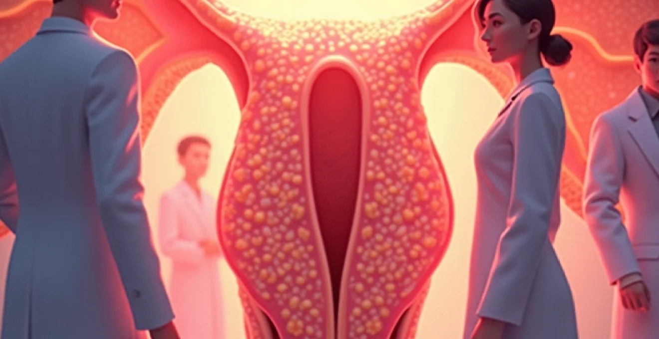
The clitoris comprises both external and internal components, forming a complex three-dimensional structure that extends deep into the pelvic cavity. While many people recognise only the external glans clitoris—the small, highly sensitive tip visible at the junction of the labia minora—the complete organ measures approximately 10 centimetres in length and resembles an inverted wishbone when viewed anatomically. This extensive structure includes the glans, shaft, crura (legs), and vestibular bulbs, all of which contribute to the organ’s remarkable sensory capabilities.
The neural distribution throughout these anatomical regions varies considerably, with the highest concentration of nerve endings found in the glans clitoris. Recent histomorphometric analysis has revealed that the dorsal nerve of the clitoris alone contains an average of 5,140 nerve fibres per side, totalling over 10,000 myelinated axons when accounting for bilateral innervation. This figure represents a conservative estimate, as it excludes unmyelinated fibres and smaller neural branches that contribute additional sensory input to the organ.
Dorsal nerve of the clitoris: primary sensory pathway
The dorsal nerve of the clitoris serves as the primary sensory conduit for this highly sensitive organ, originating from the pudendal nerve and travelling along the dorsal aspect of the clitoral shaft. These bilateral nerve structures follow a symmetrical pattern, running parallel to each other before diverging to innervate the extensive clitoral complex. The remarkable density of nerve fibres within these relatively small structures explains the intense sensations experienced during clitoral stimulation.
Research conducted through gender-affirming phalloplasty procedures has provided unprecedented access to human clitoral nerve tissue, allowing scientists to conduct precise fibre counts using advanced microscopic techniques. The specimens were magnified 1,000 times under laboratory conditions, enabling researchers to identify and quantify individual axons with remarkable precision. This methodology represents a significant advancement over previous estimates, which relied heavily on extrapolation from animal studies rather than direct human anatomical observation.
Pudendal nerve branches and clitoral innervation
The pudendal nerve system provides the foundational neural framework for clitoral sensation, branching into multiple pathways that serve different aspects of genital function. Beyond the dorsal nerve of the clitoris, additional branches innervate surrounding structures including the labia, perineum, and external anal sphincter. This interconnected network creates a complex sensory web that contributes to the multifaceted nature of sexual pleasure and genital sensitivity.
Understanding these neural connections has significant implications for surgical procedures involving the genital region. Surgeons performing labiaplasty, clitoral reconstruction, or gender-affirming procedures must navigate this intricate neural landscape carefully to preserve sensory function. The high density of nerve fibres in relatively small anatomical spaces requires extraordinary precision to avoid inadvertent damage to critical sensory pathways.
Cavernous nerves and erectile tissue connections
The clitoris contains substantial erectile tissue similar to that found in the penis, and this tissue receives innervation from cavernous nerves that regulate vascular function during sexual arousal. These autonomic nerve fibres control the complex haemodynamic changes that occur during sexual excitement, including engorgement of the clitoral body and vestibular bulbs. The coordination between sensory and autonomic neural systems creates the physiological basis for sexual response and orgasmic function.
The cavernous nerve supply to clitoral erectile tissue demonstrates the sophisticated integration of multiple neural systems in sexual function. When combined with the sensory innervation provided by the dorsal nerve, this dual neural supply creates a remarkably responsive organ capable of generating intense pleasurable sensations while simultaneously coordinating the physical changes necessary for sexual arousal and satisfaction.
Sympathetic and parasympathetic neural networks
The autonomic nervous system plays a crucial role in clitoral function through both sympathetic and parasympathetic pathways. Parasympathetic fibres, primarily originating from the sacral spinal cord segments, facilitate vasodilation and engorgement of clitoral erectile tissue during sexual arousal. Conversely, sympathetic innervation contributes to the coordination of orgasmic contractions and the return to baseline physiological states following sexual activity.
This dual autonomic innervation creates a delicate balance that enables the complex physiological responses associated with sexual pleasure. The integration of sensory input from thousands of nerve fibres with autonomic control of vascular and muscular function demonstrates the remarkable sophistication of human sexual anatomy. Understanding these neural interactions has important implications for treating sexual dysfunction and optimising surgical outcomes in reconstructive procedures.
Quantitative analysis of clitoral nerve endings
The quantification of nerve fibres in human clitoral tissue represents a significant milestone in sexual medicine research, providing the first scientifically rigorous count of neural elements in this important organ. Previous estimates of 8,000 nerve endings, widely cited in both medical literature and popular media, were based on decades-old studies conducted on bovine specimens rather than human tissue. This fundamental gap in knowledge has now been addressed through meticulous histomorphometric analysis of human clitoral nerve samples.
The research methodology employed state-of-the-art microscopic techniques and computer-assisted image analysis to achieve unprecedented accuracy in nerve fibre counting. Tissue specimens were processed using glutaraldehyde fixation, osmium tetroxide post-fixation, and embedded in araldite before sectioning into one-micrometre cross-sections. This rigorous preparation protocol ensured optimal preservation of neural structures while enabling clear visualisation of individual axons under high magnification.
The study revealed an average of 10,281 myelinated nerve fibres in the dorsal nerve of the clitoris, representing a 20% increase over previously accepted estimates and establishing a new benchmark for understanding human sexual anatomy.
Helen O’Connell’s anatomical research findings
The pioneering work of urologist Helen O’Connell fundamentally transformed our understanding of clitoral anatomy, providing the first comprehensive three-dimensional mapping of this complex organ. Her research in the late 1990s revealed that the clitoris extends far beyond the visible glans, encompassing substantial internal structures that had been largely overlooked in traditional anatomical texts. This groundbreaking work laid the foundation for subsequent neural studies by establishing the true scope and complexity of clitoral architecture.
O’Connell’s anatomical reconstructions demonstrated that the clitoris contains significantly more tissue than previously recognised, including extensive erectile structures and neural pathways. Her findings explained many aspects of female sexual response that had remained poorly understood, including the relationship between clitoral and vaginal sensation. This research established the scientific basis for recognising the clitoris as a sophisticated sensory organ rather than simply a small external structure.
Comparative nerve density studies: clitoris versus penis
Comparative analysis between clitoral and penile innervation reveals fascinating insights into the evolution and function of human sexual anatomy. While both organs develop from identical embryological tissue, their adult neural architecture displays significant differences in density and distribution patterns. The clitoris demonstrates remarkably high nerve density relative to its size, containing approximately 10,000 nerve fibres within a structure measuring just a few centimetres in length.
In contrast, the median nerve of the hand—known for its high neural density and involvement in carpal tunnel syndrome—contains approximately 18,000 nerve fibres while serving a much larger anatomical region. This comparison highlights the extraordinary concentration of sensory apparatus within the relatively small clitoral structure. Research is currently underway to quantify penile glans innervation using similar methodologies, which will provide definitive comparative data between these homologous structures.
Microscopic analysis of free nerve endings distribution
Free nerve endings represent the most abundant type of sensory receptor in clitoral tissue, providing the foundation for pain, temperature, and tactile sensation. These unmyelinated nerve terminals penetrate extensively throughout the clitoral glans and surrounding tissues, creating a dense sensory network capable of detecting subtle changes in pressure, temperature, and chemical stimuli. The distribution pattern of free nerve endings varies significantly across different regions of the clitoral complex, with the highest concentrations found in the glans region.
Advanced immunohistochemical techniques have revealed distinct populations of free nerve endings that express different neurotransmitter markers, suggesting specialised functional roles within the overall sensory apparatus. Some endings appear to be specialised for mechanical stimulation, while others respond primarily to chemical or thermal stimuli. This diversity of sensory modalities contributes to the complex and varied sensations experienced during sexual stimulation of the clitoris.
Pacinian corpuscles and meissner’s corpuscles concentration
Encapsulated nerve endings, including Pacinian corpuscles and Meissner’s corpuscles, provide specialised mechanoreceptive functions within clitoral tissue. Pacinian corpuscles respond to deep pressure and vibratory stimuli, explaining the effectiveness of vibratory stimulation in sexual arousal. These structures are distributed throughout the clitoral body and crura, with particular concentrations near the junction between the glans and shaft.
Meissner’s corpuscles, which detect light touch and low-frequency vibrations, are abundant in the superficial layers of the clitoral glans. The combination of these different mechanoreceptor types creates a sophisticated sensory system capable of discriminating between various types of tactile stimulation. This neural diversity explains why different individuals may prefer different types of touch and stimulation patterns for optimal sexual pleasure and satisfaction.
Evolutionary biology and comparative neuroanatomy
The evolutionary significance of clitoral innervation extends far beyond human sexuality, representing a fundamental aspect of mammalian reproductive biology. Comparative studies across species reveal that complex clitoral structures and associated neural networks are present in numerous mammals, suggesting strong evolutionary pressure for maintaining sophisticated genital sensory systems. The human clitoris represents the pinnacle of this evolutionary development, possessing the highest known concentration of nerve fibres relative to size among documented mammalian species.
Research has identified clitoral structures in diverse species including primates, ungulates, and even some reptiles, indicating that specialised genital sensory organs have evolved independently multiple times throughout vertebrate evolution. The presence of extensive neural networks in these structures suggests that sexual pleasure serves important biological functions beyond simple reproduction, potentially including pair bonding, stress reduction, and overall reproductive success. This evolutionary perspective provides important context for understanding the significance of human clitoral anatomy and function.
The remarkable conservation of clitoral neural architecture across mammalian species indicates that complex genital innervation provides significant adaptive advantages. Studies of primate sexuality demonstrate that species with more complex clitoral structures tend to exhibit more sophisticated social behaviours and stronger pair bonds. This correlation suggests that the evolution of dense clitoral innervation may be linked to the development of advanced social structures and reproductive strategies.
Comparative neuroanatomical studies reveal that the human clitoris possesses unique features not found in other species, including the distinctive wishbone-shaped internal structure and exceptionally high nerve density. These anatomical specialisations may reflect the unique aspects of human sexuality, including the capacity for female orgasm independent of reproduction and the importance of sexual pleasure in human pair bonding. Understanding these evolutionary adaptations provides valuable insights into both human sexual function and broader principles of mammalian reproductive biology.
Clinical implications of clitoral neural architecture
The detailed understanding of clitoral innervation has profound implications for clinical practice across multiple medical specialties. Gynaecologists, plastic surgeons, and sexual medicine specialists can now approach clitoral-related procedures with unprecedented knowledge of the neural structures they must preserve or reconstruct. This advancement has already begun to influence surgical techniques and treatment protocols, leading to improved outcomes for patients undergoing various genital procedures.
The recognition that the clitoris contains over 10,000 nerve fibres emphasises the critical importance of preserving these structures during any surgical intervention in the genital region. Even minor procedures such as episiotomies or cosmetic genital surgeries can potentially affect clitoral sensation if performed without adequate understanding of the underlying neural anatomy. This knowledge empowers surgeons to make more informed decisions about surgical approaches and techniques to minimise the risk of sensory complications.
Female genital mutilation impact on neural function
The devastating impact of female genital mutilation (FGM) on clitoral neural function can now be understood with greater precision given our enhanced knowledge of clitoral innervation. FGM procedures, which affect millions of women worldwide, cause irreversible damage to thousands of nerve fibres concentrated within the clitoral complex. The quantification of nerve density in normal clitoral tissue provides a scientific foundation for understanding the magnitude of sensory loss experienced by FGM survivors.
Reconstructive procedures for FGM survivors have evolved significantly with improved understanding of clitoral neural architecture. Surgeons can now approach clitoral reconstruction with detailed knowledge of nerve distribution patterns, enabling more precise techniques for preserving remaining neural structures while restoring anatomical form. Research into nerve regeneration and microsurgical repair techniques offers hope for partially restoring sensory function in some cases, though the complex three-dimensional architecture of clitoral innervation presents significant technical challenges.
Surgical considerations in clitoral reconstruction
Gender-affirming surgery and reconstructive procedures following trauma or congenital anomalies require detailed understanding of clitoral neural architecture to achieve optimal sensory outcomes. The identification of specific nerve populations and their distribution patterns enables surgeons to develop more sophisticated techniques for connecting appropriate neural structures during reconstructive procedures. This knowledge has already led to improvements in phalloplasty procedures, where preservation of clitoral sensation is a critical outcome measure.
Microsurgical techniques for nerve repair and reconstruction have advanced significantly with improved understanding of clitoral innervation patterns. Surgeons can now identify specific nerve branches for coaptation during reconstructive procedures, improving the likelihood of meaningful sensory recovery. The development of nerve grafting techniques and the use of conduits for bridging nerve gaps represent promising approaches for restoring function in cases of extensive neural damage.
Neurological assessment in sexual medicine
The clinical evaluation of sexual dysfunction now benefits from enhanced understanding of normal clitoral innervation patterns. Clinicians can employ more sophisticated assessment techniques to evaluate sensory function and identify specific neural pathways that may be compromised in cases of sexual dysfunction. This knowledge enables more targeted therapeutic interventions and improved diagnostic accuracy in sexual medicine practice.
Quantitative sensory testing protocols have been developed to assess different aspects of clitoral sensation, including vibration threshold, thermal sensitivity, and tactile discrimination. These assessment tools provide objective measures of neural function that can guide treatment decisions and monitor therapeutic progress. The development of standardised testing protocols based on detailed knowledge of clitoral innervation represents a significant advancement in sexual medicine diagnostics.
Contemporary research methodologies in clitoral neuroscience
Modern neuroscientific techniques have revolutionised our ability to study clitoral anatomy and function, moving beyond historical limitations imposed by cultural taboos and methodological constraints. Advanced imaging technologies, including high-resolution MRI and ultrasound, now enable detailed visualisation of clitoral structures in living individuals. These non-invasive techniques provide valuable insights into normal anatomical variation and pathological changes affecting clitoral function.
Molecular techniques have revealed the presence of specific neurotransmitter systems within clitoral neural networks, including dopaminergic, cholinergic, and nitrergic pathways. Understanding the molecular basis of clitoral sensation has opened new avenues for therapeutic intervention in cases of sexual dysfunction. Research into the role of specific receptor subtypes and signalling pathways may lead to targeted pharmacological treatments for enhancing or restoring sexual sensation.
Electrophysiological studies using sophisticated recording techniques have begun to characterise the electrical properties of clitoral nerve fibres, revealing distinct patterns of neural activity
during different types of stimulation. These studies have revealed that clitoral nerve fibres exhibit unique firing patterns that differ from those found in other sensory organs, suggesting specialised adaptations for processing sexual stimuli.The integration of computational modelling with experimental data has enabled researchers to develop sophisticated models of clitoral sensory processing. These models incorporate the complex three-dimensional anatomy of the organ along with detailed information about nerve fibre distribution and response characteristics. Such computational approaches provide valuable insights into how the remarkable density of neural elements within the clitoris contributes to its exceptional sensitivity and the generation of pleasurable sensations.Future research directions in clitoral neuroscience include the application of optogenetics techniques to selectively activate specific neural populations, potentially revealing the functional roles of different nerve fibre types within the organ. Additionally, single-cell RNA sequencing approaches may identify previously unknown neural subtypes and their specific molecular signatures. These cutting-edge methodologies promise to further enhance our understanding of this remarkable organ and its role in human sexual function.The development of standardised research protocols for studying clitoral anatomy and function represents a critical advancement in the field. International collaborations between research institutions have begun to establish consensus guidelines for tissue collection, processing, and analysis techniques. These standardised approaches will enable more robust comparisons between studies and accelerate progress in understanding clitoral neurobiology across diverse populations and clinical conditions.