The intersection of dental health and medical imaging has become increasingly relevant as magnetic resonance imaging technology advances and becomes more widespread. With over 90% of adults having experienced dental caries requiring fillings, questions about MRI safety with metallic dental restorations are more pertinent than ever. The powerful magnetic fields generated by MRI scanners can interact with various materials, creating legitimate concerns about patient safety and diagnostic accuracy. Understanding the relationship between different dental materials and magnetic field strength is crucial for both patients and healthcare professionals navigating modern medical imaging requirements.
Understanding metal dental amalgam composition and MRI magnetic field interactions
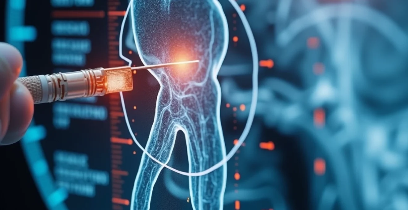
The interaction between dental materials and MRI magnetic fields depends fundamentally on the magnetic susceptibility properties of the metals involved. Modern MRI scanners generate powerful magnetic fields ranging from 1.5 Tesla to 7 Tesla, with some research facilities utilising even stronger fields. These magnetic forces are so powerful that they can move ferromagnetic objects across rooms, making safety protocols absolutely critical for patient protection.
Silver-mercury amalgam properties and ferromagnetic characteristics
Traditional amalgam fillings, commonly referred to as silver fillings due to their distinctive metallic appearance, contain a complex mixture of metals that behaves quite differently from pure ferromagnetic materials. The composition typically includes approximately 50% mercury, combined with silver, tin, copper, and trace amounts of other metals. This specific alloy combination creates a material that is essentially non-ferromagnetic , meaning it does not respond to magnetic fields in the same way that iron, cobalt, or nickel would.
Research has consistently demonstrated that standard-strength MRI scanners (1.5T and 3T) do not pose safety risks to patients with amalgam fillings. The magnetic field strength at these levels is insufficient to create dangerous heating, movement, or displacement of properly set amalgam restorations. However, recent studies have raised concerns about ultra-high-field MRI scanners operating at 7 Tesla, where mercury leakage from amalgam fillings has been observed under laboratory conditions.
Gold alloy inlays and their diamagnetic response to tesla field strength
Gold-based dental restorations represent another category of metallic dental work that patients frequently encounter. Pure gold is classified as a diamagnetic material , meaning it creates a weak repulsive force when exposed to magnetic fields. This property makes gold dental work generally safe for MRI procedures, though the material can still create imaging artefacts that may compromise diagnostic quality.
Gold alloys used in dentistry often contain small amounts of other metals to improve durability and workability. These additions can slightly alter the magnetic properties, but rarely to a degree that creates safety concerns. The primary consideration with gold dental work during MRI scans relates to image quality rather than patient safety, as the metal can cause signal dropout and geometric distortion in the immediate vicinity of the restoration.
Titanium implant materials and paramagnetic behaviour in 1.5T and 3T scanners
Titanium has become the gold standard for dental implants due to its exceptional biocompatibility and paramagnetic properties . Unlike ferromagnetic materials that are strongly attracted to magnetic fields, titanium exhibits only weak attraction that poses no safety risks during MRI procedures. The material’s paramagnetic nature means it can cause minor image distortions, but these are typically minimal compared to other metallic dental materials.
Modern titanium dental implants undergo rigorous testing to ensure MRI compatibility. The manufacturing processes and alloy compositions are specifically designed to minimise magnetic susceptibility while maintaining the structural integrity required for long-term dental function. Patients with titanium implants can generally undergo MRI scans without special precautions, though radiologists should be informed of their presence to optimise imaging protocols.
Stainless steel crown components and magnetic susceptibility artefacts
Older dental restorations, particularly those placed before the 1990s, may contain stainless steel components that exhibit stronger magnetic properties. Stainless steel contains varying amounts of iron and other ferromagnetic elements, making it potentially problematic during MRI procedures. The specific grade and composition of stainless steel used in dental applications can significantly influence its behaviour in magnetic fields.
Patients with older crowns, bridges, or orthodontic appliances should inform their healthcare providers about the age and type of their dental work. In some cases, dental records may need to be reviewed to determine the exact materials used. While complete removal of stainless steel dental work is rarely necessary, alternative imaging sequences or protocols may be recommended to ensure both safety and diagnostic quality.
MRI safety protocols for patients with metallic dental restorations
Comprehensive safety protocols have been established to protect patients with metallic dental restorations during MRI procedures. These protocols involve multiple checkpoints and assessment procedures designed to identify potential risks before patients enter the MRI environment. The systematic approach ensures that both obvious and subtle safety concerns are addressed appropriately.
Pre-screening questionnaire requirements and dental history documentation
Every patient scheduled for an MRI must complete a detailed pre-screening questionnaire that includes specific questions about dental history and current dental work. This questionnaire serves as the first line of defence against potential complications, requiring patients to disclose information about fillings, crowns, bridges, implants, and orthodontic appliances. The questionnaire also asks about the approximate dates when dental work was completed, as this information helps determine the likely materials used.
Accurate completion of pre-screening documentation requires patients to be honest about their dental history, even if they are uncertain about specific materials or dates. Healthcare providers emphasise that no judgment is made about dental care choices, and the information is used solely for safety purposes. Patients should contact their dentists if they need clarification about their dental restorations before their MRI appointment.
Radiological technologist assessment procedures for metal detection
Qualified radiological technologists conduct thorough assessments of patients before MRI procedures, reviewing pre-screening questionnaires and asking follow-up questions as needed. These professionals are trained to identify potential risk factors and make appropriate decisions about imaging protocols. The assessment may include visual examination of visible dental work and consultation with radiologists or referring physicians when unusual circumstances arise.
Some facilities utilise handheld metal detectors or walk-through metal detection systems as additional screening measures. These tools can identify metallic objects that patients may have forgotten to mention or may not have realised contained metal. The detection systems are particularly useful for identifying smaller metallic items that might otherwise be overlooked during visual inspection.
Contraindication guidelines for ferromagnetic dental materials
Specific contraindication guidelines exist for patients with known ferromagnetic dental materials, though absolute contraindications are relatively rare in modern dental practice. The guidelines are based on extensive research and clinical experience, taking into account factors such as magnetic field strength, imaging sequences planned, and the location of metallic objects relative to the area being imaged.
Patients with ferromagnetic dental materials may require modified imaging protocols or alternative diagnostic approaches, but complete exclusion from MRI is uncommon when proper safety measures are followed.
When contraindications are identified, healthcare providers work with patients to explore alternative imaging options or modifications to the planned MRI procedure. These alternatives might include different imaging sequences, reduced magnetic field strength, or completely different imaging modalities such as CT scanning or ultrasound, depending on the diagnostic requirements.
Patient positioning modifications to minimise susceptibility artefacts
Strategic patient positioning can significantly reduce the impact of metallic dental restorations on image quality while maintaining safety standards. Radiological technologists may adjust head positioning, coil placement, or imaging angles to minimise the influence of dental work on the areas of primary diagnostic interest. These modifications require expertise in both MRI physics and anatomy to ensure optimal results.
Positioning modifications are particularly important when imaging the head, neck, or upper spine, where dental work is most likely to create problematic artefacts. The technologist may use specialised cushioning, head restraints, or positioning aids to achieve the optimal angle for imaging while ensuring patient comfort throughout the procedure.
Image quality degradation and artefact formation mechanisms
The presence of metallic dental restorations can significantly impact MRI image quality through various physical mechanisms. Understanding these artefact formation processes helps both healthcare providers and patients appreciate why certain modifications to imaging protocols may be necessary. The severity and type of artefacts depend on multiple factors including metal composition, restoration size, magnetic field strength, and specific imaging sequences used.
Magnetic susceptibility artefacts in T1-Weighted and T2-Weighted sequences
Magnetic susceptibility artefacts represent the most common type of image degradation caused by metallic dental work. These artefacts appear as areas of signal dropout, geometric distortion, or abnormal signal intensity surrounding metallic objects. T1-weighted sequences typically show these artefacts as areas of decreased signal intensity, while T2-weighted sequences may demonstrate more complex patterns of signal alteration.
The extent of susceptibility artefacts is directly related to the magnetic properties of the dental materials and the strength of the MRI magnetic field. Materials with higher magnetic susceptibility create more pronounced artefacts, potentially obscuring anatomical structures or creating false impressions of pathology. Radiologists must carefully distinguish between artefact-related findings and genuine pathological conditions when interpreting images from patients with metallic dental work.
Signal dropout patterns around amalgam fillings in FLAIR imaging
Fluid Attenuated Inversion Recovery (FLAIR) imaging sequences are particularly sensitive to the presence of amalgam fillings, often showing characteristic signal dropout patterns in adjacent brain tissue. These patterns can create diagnostic challenges when evaluating conditions such as multiple sclerosis, stroke, or brain tumours, where subtle signal changes may be clinically significant.
The signal dropout typically appears as dark areas extending several millimetres around amalgam fillings, with the extent varying based on the size and number of restorations present. Radiologists experienced in interpreting images from patients with dental work can often distinguish between artefact-related signal changes and genuine pathology, but additional imaging may sometimes be necessary for definitive diagnosis.
Geometric distortion effects on Diffusion-Weighted imaging protocols
Diffusion-weighted imaging (DWI) sequences are particularly vulnerable to geometric distortions caused by metallic dental restorations. These distortions can significantly impact the assessment of brain tissue integrity, stroke evaluation, and other conditions where precise anatomical localisation is crucial. The distortions typically manifest as stretching, compression, or displacement of anatomical structures in the vicinity of metallic objects.
Advanced DWI protocols may incorporate distortion correction algorithms or alternative acquisition techniques to minimise the impact of metallic dental work. However, these corrections have limitations, and some degree of image degradation may persist despite technical optimisation. Radiologists must account for these limitations when interpreting DWI studies from patients with extensive dental work.
Gradient echo sequence vulnerabilities to Metal-Induced field inhomogeneities
Gradient echo sequences demonstrate particular sensitivity to magnetic field inhomogeneities created by metallic dental restorations. These sequences rely on precise magnetic field uniformity to generate high-quality images, making them vulnerable to even small amounts of metallic material. The resulting artefacts can appear as severe signal dropout, geometric distortion, or complete loss of diagnostic information in affected areas.
The vulnerability of gradient echo sequences to metal-induced artefacts has led to the development of alternative imaging approaches for patients with extensive dental work. Spin echo sequences, which are more robust in the presence of field inhomogeneities, may be substituted when gradient echo imaging is compromised by dental metalwork.
Clinical case studies and Evidence-Based safety data
Extensive clinical research and case study analysis have provided substantial evidence regarding MRI safety for patients with metallic dental restorations. Large-scale studies involving thousands of patients have consistently demonstrated that conventional MRI scanning at 1.5T and 3T field strengths poses minimal safety risks for individuals with standard amalgam fillings, gold crowns, and titanium implants.
A comprehensive analysis of adverse events related to dental work during MRI procedures found that serious complications are extraordinarily rare, occurring in fewer than 0.01% of scanned patients. The few reported incidents typically involved older dental work containing materials not commonly used in modern dentistry, or situations where pre-screening protocols were not properly followed.
Recent research has raised concerns specifically about ultra-high-field 7 Tesla MRI scanners and their interaction with amalgam fillings. Laboratory studies using extracted teeth demonstrated measurable mercury release when amalgam fillings were exposed to 7T magnetic fields for extended periods. However, the clinical significance of this finding remains unclear, as the experimental conditions did not perfectly replicate real-world scanning scenarios.
Clinical evidence suggests that while ultra-high-field MRI may cause mercury release from amalgam fillings under laboratory conditions, the practical health implications for patients remain uncertain and require further investigation.
Long-term follow-up studies of patients who have undergone multiple MRI scans with metallic dental work show no increased incidence of dental complications, oral health problems, or systemic health issues attributable to MRI exposure. These findings provide reassurance about the safety of routine MRI scanning for patients with conventional dental restorations.
Advanced MRI techniques for metal artefact reduction
The development of sophisticated metal artefact reduction techniques has significantly improved imaging capabilities for patients with metallic dental restorations. These advanced methods employ various strategies to minimise the impact of dental work on image quality while maintaining diagnostic accuracy. The techniques range from simple protocol modifications to complex post-processing algorithms that can substantially reduce artefact severity.
View angle tilting (VAT) represents one of the most effective techniques for reducing metal-induced geometric distortions. This method adjusts the readout gradient during image acquisition to counteract the distorting effects of magnetic field inhomogeneities around metallic objects. VAT is particularly effective for reducing the stretching and compression artefacts commonly seen with dental metalwork.
Slice encoding for metal artefact correction (SEMAC) and multi-acquisition variable-resonance image combination (MAVRIC) represent more advanced approaches that acquire multiple images with different encoding parameters. These techniques then combine the images using sophisticated algorithms to create final images with dramatically reduced metal artefacts. The methods require longer scan times but can produce diagnostic-quality images even in the presence of extensive metalwork.
Short tau inversion recovery (STIR) sequences offer another approach for imaging patients with metallic dental restorations. These sequences are inherently more robust in the presence of magnetic field inhomogeneities and often provide better image quality around metallic objects compared to conventional sequences. STIR imaging is particularly valuable for evaluating soft tissues adjacent to dental work.
Alternative imaging modalities for patients with extensive dental metalwork
When MRI imaging is significantly compromised by extensive metallic dental work, alternative imaging modalities may provide better diagnostic information. Computed tomography (CT) scanning, while involving ionising radiation exposure, is largely unaffected by dental metalwork and can provide excellent anatomical detail for many clinical applications. Modern CT scanners incorporate metal artefact reduction algorithms that minimise streak artefacts from dental restorations.
Ultrasound imaging represents a radiation-free alternative that can be valuable for evaluating certain conditions affecting the head and neck region. High-resolution ultrasound can assess soft tissue structures, blood flow, and some pathological conditions without interference from dental metalwork. However, ultrasound has limitations in evaluating deep structures and bony anatomy.
Positron emission tomography (PET) imaging, often combined with CT, provides functional information about tissue metabolism that complements anatomical imaging. PET imaging is generally unaffected by dental metalwork, though the CT component may show some artefacts. This modality can be particularly valuable for evaluating certain types of pathology where functional information is more important than fine anatomical detail.
The choice between alternative imaging modalities depends on the specific clinical question being addressed, the extent of dental metalwork present, and individual patient factors such as radiation sensitivity or contrast agent allergies. Healthcare providers work closely with patients to determine the most appropriate imaging approach for each unique situation, ensuring that diagnostic needs are met while minimising any potential risks or discomfort.