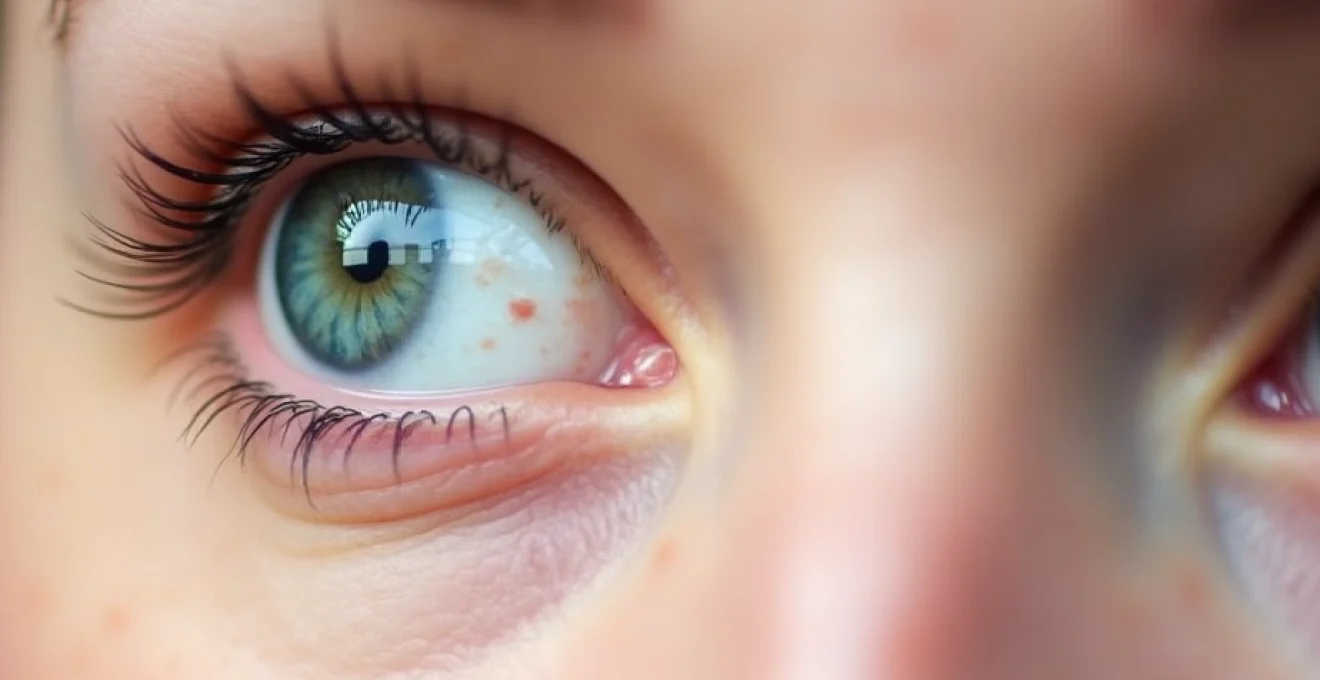
Left eyelid twitching, medically known as myokymia, represents one of the most common yet misunderstood neurological phenomena affecting millions of individuals worldwide. This involuntary muscle contraction of the orbicularis oculi muscle can range from barely perceptible tremors to pronounced spasms that significantly impact daily activities. While most cases prove benign and self-limiting, understanding the underlying mechanisms, potential causes, and appropriate treatment strategies becomes essential for both healthcare professionals and patients experiencing this condition.
The prevalence of eyelid fasciculations has increased dramatically in recent years, with approximately 70% of the population experiencing some form of periorbital muscle spasm during their lifetime. This surge correlates strongly with modern lifestyle factors, including increased screen time, elevated stress levels, and dietary deficiencies that characterise contemporary society. The complexity of eyelid twitching extends beyond simple muscle fatigue, encompassing intricate neurological pathways, biochemical imbalances, and potentially serious underlying pathological conditions .
Understanding left eyelid twitching: myokymia and fasciculation mechanisms
Orbicularis oculi muscle anatomy and neural innervation
The orbicularis oculi muscle consists of three distinct anatomical components: the orbital, palpebral, and lacrimal portions. Each segment serves specific functions in eyelid closure, blinking reflexes, and tear drainage mechanisms. The orbital portion provides forceful eyelid closure during sleep and protective reflexes, while the palpebral portion enables gentle blinking and voluntary eyelid movements. Neural innervation occurs primarily through the facial nerve (cranial nerve VII), specifically the temporal and zygomatic branches that supply motor function to the periorbital musculature.
The sophisticated neural circuitry controlling eyelid movement involves multiple levels of the central nervous system, from cortical motor areas to brainstem nuclei. The facial nucleus in the pons serves as the primary motor centre, receiving inputs from higher cortical areas, the reticular formation, and sensory feedback loops. Disruption at any level of this complex pathway can manifest as abnormal muscle contractions, ranging from subtle fasciculations to severe blepharospasm .
Benign essential blepharospasm vs simple eyelid myokymia
Distinguishing between benign eyelid myokymia and essential blepharospasm proves crucial for appropriate management and prognosis. Simple myokymia typically presents as fine, rippling contractions affecting small muscle bundles within the orbicularis oculi. These contractions remain localised, rarely spreading to adjacent facial muscles, and generally resolve spontaneously within days to weeks. The movements appear subtle to external observers and cause minimal functional impairment.
Essential blepharospasm, conversely, represents a more severe dystonic disorder characterised by sustained, involuntary contractions that force eyelid closure. This condition affects both upper and lower eyelids simultaneously, often spreading to involve adjacent facial muscles in a pattern known as Meige syndrome. Patients with essential blepharospasm experience significant functional disability, including reading difficulties, driving impairment, and social embarrassment due to the visible nature of the contractions .
Facial nerve VII motor unit hyperexcitability patterns
Facial nerve hyperexcitability represents a key pathophysiological mechanism underlying many forms of eyelid twitching. This phenomenon occurs when motor units within the facial nerve demonstrate increased responsiveness to normal stimuli or generate spontaneous activity in the absence of voluntary commands. Several factors contribute to this hyperexcitability, including mechanical compression of nerve fibres, metabolic disturbances affecting nerve membrane stability, and central nervous system lesions affecting inhibitory control mechanisms.
Research has identified specific patterns of motor unit recruitment during eyelid fasciculations. High-frequency, low-amplitude discharges characterise benign myokymia, while essential blepharospasm demonstrates more complex patterns involving multiple motor unit synchronisation.
Understanding these electrophysiological signatures enables clinicians to differentiate between various forms of eyelid twitching and guide appropriate therapeutic interventions
.
Electromyographic characteristics of periorbital muscle spasms
Electromyographic analysis provides valuable insights into the nature and severity of periorbital muscle spasms. Surface EMG recordings from the orbicularis oculi typically reveal characteristic burst patterns during fasciculation episodes. Benign myokymia demonstrates brief, irregular bursts lasting 100-200 milliseconds, with frequencies ranging from 3-8 Hz. These bursts occur sporadically, with quiet periods lasting several seconds to minutes between episodes.
More severe forms of blepharospasm exhibit prolonged bursts lasting several seconds, often accompanied by co-contraction of antagonist muscles. The EMG patterns may show synchronisation between different portions of the orbicularis oculi, indicating central nervous system involvement rather than peripheral nerve irritation. Needle EMG examination can reveal additional abnormalities, including increased insertional activity, positive sharp waves, and abnormal spontaneous activity suggestive of nerve damage or muscle denervation .
Primary triggers and underlying pathophysiology of left eyelid spasms
Caffeine-induced adenosine receptor antagonism effects
Caffeine consumption represents one of the most prevalent triggers for eyelid fasciculations, acting through competitive antagonism of adenosine receptors in the central nervous system. Adenosine normally functions as an inhibitory neurotransmitter, promoting muscle relaxation and neural circuit dampening. When caffeine blocks these receptors, it eliminates this inhibitory influence, leading to increased neural excitability and heightened muscle responsiveness to stimuli.
The dose-response relationship between caffeine intake and eyelid twitching demonstrates significant individual variation. Some individuals experience fasciculations with as little as 100mg of caffeine (equivalent to one cup of coffee), while others tolerate much higher doses without symptoms. Chronic caffeine consumption leads to adenosine receptor upregulation, requiring progressively higher doses to achieve the same stimulating effects while simultaneously increasing susceptibility to withdrawal-related muscle spasms .
Sleep deprivation and circadian rhythm disruption impact
Sleep deprivation profoundly affects neuromuscular function through multiple pathways, making it a significant risk factor for eyelid fasciculations. During normal sleep, the body undergoes essential restorative processes, including neurotransmitter replenishment, metabolic waste clearance from brain tissues, and muscle recovery. Insufficient sleep disrupts these processes, leading to accumulated cellular damage and impaired neural function.
Circadian rhythm disruption, common in shift workers and individuals with irregular sleep schedules, compounds these effects by desynchronising internal biological clocks. This desynchronisation affects hormone production, including cortisol and melatonin, which play crucial roles in muscle tone regulation and neural excitability. Research indicates that individuals sleeping less than six hours per night show a three-fold increase in eyelid fasciculation frequency compared to those maintaining regular eight-hour sleep schedules.
Chronic stress and Cortisol-Mediated neuromuscular hyperactivity
Chronic psychological stress triggers a cascade of physiological responses that significantly impact neuromuscular function. The hypothalamic-pituitary-adrenal axis becomes overactive during prolonged stress, leading to excessive cortisol production. Elevated cortisol levels affect muscle membrane stability, alter neurotransmitter metabolism, and increase overall neural excitability throughout the nervous system.
Stress-related eyelid twitching often accompanies other somatic symptoms, including muscle tension headaches, jaw clenching, and generalised muscle stiffness. The condition frequently worsens during periods of high psychological demand, such as examination periods, work deadlines, or major life transitions.
Clinical observations suggest that stress-induced fasciculations respond well to comprehensive stress management approaches, including relaxation techniques, cognitive behavioural therapy, and lifestyle modifications
.
Digital eye strain and blue light exposure consequences
The digital revolution has introduced new challenges for ocular health, with prolonged screen exposure contributing significantly to eyelid fasciculation development. Digital eye strain, also known as computer vision syndrome, affects up to 90% of individuals who spend more than three hours daily using digital devices. The condition results from reduced blink rates during screen use, leading to ocular surface desiccation and compensatory muscle hyperactivity.
Blue light emission from digital screens may contribute to fasciculation development through disruption of circadian rhythms and increased oxidative stress in retinal tissues. Studies demonstrate that blue light exposure in the evening hours suppresses melatonin production, affecting sleep quality and indirectly contributing to muscle hyperexcitability. The implementation of blue light filters and regular screen breaks can significantly reduce the incidence of screen-related eyelid twitching .
Magnesium deficiency and calcium channel dysfunction
Magnesium deficiency represents a frequently overlooked cause of muscle fasciculations, including eyelid twitching. This essential mineral serves as a natural calcium channel blocker, regulating muscle contraction and neural transmission. When magnesium levels become insufficient, calcium channels remain inappropriately open, leading to excessive muscle contractions and increased neural excitability.
Modern dietary patterns often provide insufficient magnesium intake, with processed foods containing significantly lower levels compared to whole, unprocessed alternatives. Factors that increase magnesium requirements include physical stress, alcohol consumption, certain medications, and underlying medical conditions such as diabetes or gastrointestinal disorders. Laboratory assessment typically reveals serum magnesium levels below 1.8 mg/dL in individuals with deficiency-related fasciculations, though intracellular magnesium depletion can occur despite normal serum levels.
Neurological conditions associated with unilateral eyelid tremor
Hemifacial spasm and vascular compression syndromes
Hemifacial spasm presents as unilateral facial muscle contractions that typically begin with eyelid twitching before progressing to involve the entire ipsilateral facial musculature. This condition results from vascular compression of the facial nerve at its root exit zone from the brainstem, most commonly by the anterior inferior cerebellar artery or posterior inferior cerebellar artery. The compression creates ephaptic transmission between nerve fibres, leading to abnormal synchronisation of motor unit activity.
The natural history of hemifacial spasm demonstrates progressive worsening over time, with initial subtle eyelid contractions evolving into pronounced facial distortions that significantly impact quality of life. Patients often report social embarrassment, functional limitations in activities requiring fine facial control, and psychological distress related to the visible nature of the contractions. Early recognition becomes crucial, as timely intervention with botulinum toxin injections or microvascular decompression surgery can provide substantial symptom relief .
Bell’s palsy recovery phase synkinesis development
Following Bell’s palsy, approximately 30% of patients develop synkinetic movements during the recovery phase, where intended movements of one facial muscle group trigger involuntary contractions in other areas. This phenomenon occurs due to aberrant reinnervation of facial muscles by regenerating nerve fibres that form inappropriate connections with their target tissues. Eyelid-oral synkinesis represents a common pattern, where attempts to smile or speak trigger involuntary eyelid closure or twitching.
The development of post-Bell’s palsy synkinesis typically begins 3-6 months after the initial paralysis, coinciding with nerve regeneration processes. Early signs include subtle twitching movements that occur synchronously with voluntary facial expressions. Over time, these movements may become more pronounced and functionally limiting. Physical therapy focusing on facial muscle re-education and selective botulinum toxin injections can help minimise synkinetic patterns and restore more normal facial function.
Multiple sclerosis demyelination and cranial nerve involvement
Multiple sclerosis can affect the facial nerve pathways at various locations, from the brainstem nuclei to peripheral nerve segments. Demyelinating lesions disrupt normal conduction patterns, leading to abnormal spontaneous activity and increased excitability of facial motor neurons. Eyelid fasciculations in multiple sclerosis patients may represent early manifestations of brainstem involvement, often preceding more obvious neurological deficits.
The characteristics of multiple sclerosis-related eyelid twitching differ from benign fasciculations in several important ways. The contractions tend to be more persistent, may involve both eyelids simultaneously, and often occur in conjunction with other neurological symptoms such as diplopia, facial numbness, or speech difficulties.
Magnetic resonance imaging frequently reveals characteristic demyelinating lesions in the brainstem or cerebellum, helping to establish the diagnosis and guide appropriate disease-modifying therapies
.
Dystonia and basal ganglia movement disorder manifestations
Dystonic disorders affecting the periorbital region can present initially as seemingly benign eyelid twitching before progressing to more characteristic sustained contractions. These conditions result from dysfunction within basal ganglia circuits responsible for movement control and inhibition. Primary dystonia typically begins in adulthood and affects women more frequently than men, with a characteristic age of onset between 40-60 years.
The progression from simple fasciculations to frank dystonia occurs gradually over months to years. Early signs include increased frequency and duration of contractions, spreading to involve adjacent muscle groups, and development of sensory tricks (geste antagoniste) that temporarily relieve symptoms. Environmental factors such as bright lights, wind, or emotional stress often exacerbate symptoms, while concentrating on specific tasks may temporarily suppress the movements. Genetic testing may reveal mutations in genes associated with dystonia, including DYT1, DYT5, and DYT12, particularly in cases with early onset or family history .
Pharmaceutical and toxicological causes of periorbital fasciculations
Numerous medications can trigger or exacerbate eyelid fasciculations through various mechanisms affecting neuromuscular transmission. Stimulant medications, including amphetamines, methylphenidate, and modafinil, increase dopaminergic and noradrenergic activity, leading to heightened muscle excitability. Antidepressants, particularly selective serotonin reuptake inhibitors, can induce fasciculations through serotonergic modulation of motor neuron excitability.
Topical ophthalmic medications present unique risks for periorbital fasciculations due to their direct application near target tissues. Beta-agonist bronchodilators, commonly used in asthma inhalers, can cause systemic absorption leading to tremor and fasciculations. Lithium therapy, used in bipolar disorder treatment, frequently causes fine tremor and muscle fasciculations through effects on sodium channel function and neurotransmitter metabolism.
Environmental toxin exposure represents an increasingly recognised cause of neuromuscular symptoms, including eyelid twitching. Organophosphate pesticides inhibit acetylcholinesterase, leading to excessive cholinergic stimulation and muscle hyperactivity. Heavy metal poisoning, particularly mercury and lead exposure, can cause widespread fasciculations as part of broader neurotoxicity syndromes. Occupational exposure assessment becomes essential in patients with unexplained fasciculations, especially those working in agriculture, manufacturing, or other high-risk industries .
Evidence-based treatment protocols and therapeutic interventions
Botulinum toxin type A injection techniques for blepharospasm
Botulinum toxin type A injections represent the gold standard treatment for severe eyelid fasciculations and blepharospasm. The neurotoxin works by blocking acetylcholine release at neuromuscular junctions, effectively paralysing overactive muscle fibres. Precise injection technique proves critical for optimal outcomes while minimising complications such as ptosis, diplopia, or dry eye syndrome.
Standard injection protocols involve multiple small aliquots distributed across the orbicularis oculi muscle, with typical doses ranging from 12-30 units per eye depending on symptom severity and patient response. The lateral portions of the upper and lower eyelids receive priority treatment, as these areas typically demonstrate the most pronounced contractions. Treatment effects typically begin within 3-7 days, reach maximum benefit at 2-4 weeks, and maintain efficacy for 3-6 months before requiring repeat injections.
Recent advances in injection techniques include the use of electromyographic guidance for precise muscle targeting and the development of novel botulinum tox
in formulations that provide longer-lasting effects. Patient selection criteria include failed conservative management, functional impairment affecting daily activities, and absence of contraindications such as neuromuscular disorders or pregnancy.
Magnesium supplementation and electrolyte rebalancing strategies
Magnesium supplementation forms the cornerstone of treatment for deficiency-related eyelid fasciculations, with various formulations offering different bioavailability profiles. Magnesium glycinate and magnesium taurate demonstrate superior absorption compared to magnesium oxide, with fewer gastrointestinal side effects. Standard dosing typically ranges from 200-400mg daily, divided into two doses to optimise absorption and minimise digestive upset.
Laboratory monitoring becomes essential during supplementation, as excessive magnesium intake can lead to hypermagnesemia and associated complications. Serum magnesium levels should be assessed before initiating therapy and monitored periodically during treatment. Concurrent evaluation of other electrolytes, including potassium, calcium, and phosphorus, helps identify additional imbalances that may contribute to muscle hyperexcitability. Patients with kidney disease require careful dose adjustments, as impaired renal function can lead to magnesium accumulation and toxicity.
Stress reduction techniques and parasympathetic nervous system activation
Comprehensive stress management protocols target the underlying sympathetic hyperactivation that contributes to eyelid fasciculations. Progressive muscle relaxation techniques teach patients to systematically tense and release muscle groups, promoting awareness of muscle tension patterns and facilitating voluntary relaxation. Deep breathing exercises activate the parasympathetic nervous system, counteracting stress-induced muscle hyperexcitability through vagal stimulation.
Mindfulness-based interventions demonstrate particular efficacy in reducing stress-related fasciculations. Regular meditation practice alters brain activity patterns in regions associated with stress response, including the amygdala and prefrontal cortex. Biofeedback therapy provides real-time monitoring of physiological parameters, enabling patients to develop conscious control over autonomic nervous system responses. Research indicates that individuals practicing stress reduction techniques experience a 60-70% reduction in fasciculation frequency within 4-6 weeks of consistent implementation.
The integration of cognitive behavioural therapy with physiological stress reduction techniques provides optimal outcomes for patients with stress-induced eyelid fasciculations, addressing both psychological triggers and physical manifestations
Ophthalmological lubricant therapy for dry eye syndrome
Artificial tear therapy addresses the ocular surface dysfunction that frequently accompanies eyelid fasciculations, particularly in cases related to digital eye strain or environmental factors. Preservative-free formulations prove superior for frequent use, as preserved drops can cause additional irritation and exacerbate symptoms. Hyaluronic acid-based lubricants provide longer-lasting surface protection compared to traditional polyvinyl alcohol formulations.
Treatment protocols typically involve frequent application during symptomatic periods, with maintenance therapy continuing even after fasciculations resolve. Lipid-based artificial tears benefit patients with meibomian gland dysfunction, while gel formulations provide enhanced lubrication for severe dry eye cases. Environmental modifications, including humidifier use and positioning adjustments relative to air conditioning vents, complement topical therapy. Patients should avoid over-the-counter vasoconstrictive eye drops, which can worsen dry eye symptoms and potentially trigger rebound fasciculations.
When to seek medical evaluation: red flag symptoms and differential diagnosis
While most eyelid fasciculations represent benign, self-limiting conditions, certain clinical features warrant immediate medical evaluation. Progressive weakness of eyelid closure, spreading of fasciculations to other facial muscles, or development of sustained eyelid spasms suggest more serious underlying pathology. Concurrent neurological symptoms, including speech difficulties, swallowing problems, or limb weakness, may indicate central nervous system involvement requiring urgent investigation.
The presence of ptosis, particularly if asymmetric or progressive, raises concern for neuromuscular junction disorders such as myasthenia gravis. Diplopia accompanying eyelid fasciculations suggests cranial nerve involvement or brainstem pathology. Visual field defects, altered pupillary responses, or changes in visual acuity require ophthalmological evaluation to exclude structural abnormalities or increased intracranial pressure. Patients experiencing fasciculations lasting more than three weeks, or those with family history of movement disorders, should undergo neurological assessment to exclude hereditary conditions.
Comprehensive clinical evaluation includes detailed medication history, occupational exposure assessment, and systematic neurological examination. Laboratory studies may include complete blood count, comprehensive metabolic panel, thyroid function tests, and vitamin B12 levels. Electromyographic studies help differentiate between peripheral nerve disorders and central nervous system pathology. Advanced imaging, including brain MRI, becomes necessary when clinical features suggest structural abnormalities or demyelinating disease. Have you noticed any changes in your symptoms that might indicate progression beyond simple fasciculations? Early recognition and appropriate referral can significantly impact treatment outcomes and prevent potential complications from undiagnosed neurological conditions.