White patches appearing on fingernails represent one of the most frequently encountered nail abnormalities in clinical practice, affecting millions of individuals worldwide regardless of age or gender. These distinctive discolourations, medically termed leukonychia, can manifest as tiny pinpoint spots, horizontal bands, or complete nail whitening. While often dismissed as merely cosmetic concerns, white nail patches may occasionally signal underlying systemic conditions ranging from nutritional deficiencies to serious organ dysfunction. Understanding the various causes behind nail leukonychia enables both healthcare professionals and individuals to distinguish between benign trauma-related changes and potentially significant health indicators requiring medical attention.
Leukonychia: medical classification and pathophysiology
Leukonychia encompasses a diverse spectrum of nail plate discolourations characterised by the presence of white areas within the otherwise translucent nail structure. The condition derives its name from the Greek words “leukos” meaning white and “onyx” meaning nail, providing a straightforward description of this common nail manifestation. Medical professionals classify leukonychia based on several criteria including distribution patterns, underlying pathophysiology, and associated clinical presentations.
The fundamental mechanism behind leukonychia involves disruption of normal nail plate formation at the level of the nail matrix, where keratinocytes undergo differentiation to form the nail plate. When this process becomes compromised, air pockets or altered keratin structures create optical changes that appear as white areas. The nail matrix, located beneath the proximal nail fold, serves as the primary site of nail production and represents the most vulnerable area to various pathological processes.
Leukonychia punctata: isolated white spot formation
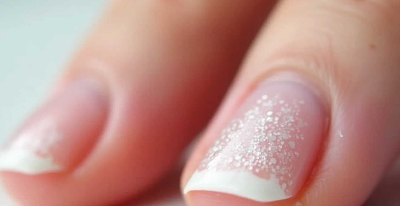
Punctate leukonychia manifests as small, isolated white spots scattered across the nail plate surface. These circular or oval lesions typically measure between 1-3 millimetres in diameter and represent the most common form of nail leukonychia encountered in clinical practice. The spots develop following localised trauma to specific areas of the nail matrix, creating microscopic air bubbles within the developing nail plate structure.
Research indicates that punctate leukonychia affects approximately 40% of the general population at some point during their lifetime. The condition demonstrates no particular gender predisposition and can occur across all age groups, though it appears more frequently in children and young adults due to increased likelihood of nail trauma during recreational activities.
Leukonychia striata: transverse white line development
Striata leukonychia presents as horizontal white bands extending across the nail plate width, typically running parallel to the lunula. These bands, also known as Mees’ lines when associated with systemic conditions, develop when the entire width of the nail matrix experiences simultaneous disruption. The resulting white lines move distally as the nail grows, eventually disappearing at the free edge.
The formation of transverse white lines often correlates with systemic stressors affecting nail matrix function uniformly across its width. This pattern suggests that the underlying cause impacted nail production globally rather than in isolated areas, distinguishing it from the localised trauma patterns seen in punctate leukonychia.
Leukonychia totalis: complete nail plate discolouration
Total leukonychia represents the most dramatic form of nail whitening, involving complete discolouration of the entire nail plate. This rare condition affects less than 1% of the population and typically indicates either congenital abnormalities or severe systemic disease processes. The uniformly white appearance results from extensive disruption of normal nail matrix function affecting the entire nail production apparatus.
Hereditary forms of total leukonychia often manifest during early childhood and may be associated with specific genetic mutations affecting keratin production. Acquired total leukonychia , conversely, usually develops in adulthood and frequently accompanies serious medical conditions requiring immediate clinical evaluation and management.
Leukonychia partialis: partial nail matrix involvement
Partial leukonychia encompasses all forms of incomplete nail whitening, including punctate and striata variants as well as irregular patterns not fitting specific categories. This classification system helps clinicians organise their diagnostic approach and determine appropriate investigation strategies based on the extent and pattern of nail involvement.
The clinical significance of partial leukonychia varies considerably depending on the underlying cause and associated symptoms. While isolated nail changes often represent benign conditions, the presence of multiple affected nails or accompanying systemic symptoms may warrant comprehensive medical evaluation to exclude underlying pathology.
Traumatic nail matrix injury and keratinocyte disruption
Physical trauma represents the most prevalent cause of leukonychia, accounting for approximately 80% of all white nail spot occurrences in healthy individuals. The nail matrix demonstrates particular vulnerability to mechanical injury due to its location beneath the proximal nail fold and its role as the active site of nail production. Even minor trauma can disrupt the delicate process of keratinocyte differentiation, leading to visible white patches that become apparent weeks after the initial injury.
The temporal relationship between trauma and visible leukonychia often creates diagnostic confusion, as patients rarely associate current nail changes with injuries that occurred several weeks previously. Fingernails grow at approximately 3-4 millimetres per month, meaning white spots may not become visible until 6-8 weeks following the initial matrix injury. This delayed manifestation frequently leads to misattribution of nail changes to recent activities rather than the actual causative event.
Mechanical trauma from manicure procedures
Professional and home manicure procedures represent significant sources of nail matrix trauma, particularly when aggressive cuticle manipulation or excessive nail filing techniques are employed. The use of metal instruments near the nail matrix can create microscopic injuries that subsequently manifest as white spots or lines. Overzealous cuticle cutting poses particular risk, as the instruments may inadvertently damage the underlying matrix tissue.
Studies examining manicure-related nail trauma suggest that approximately 15-20% of regular manicure recipients develop some form of leukonychia within six months of beginning regular treatments. The risk increases substantially when non-professional services are utilised or when proper sterilisation protocols are not followed, leading to both traumatic and infectious causes of nail discolouration.
Onychophagia-related matrix damage
Chronic nail biting, medically termed onychophagia, creates repetitive trauma to both the nail plate and surrounding matrix tissue. This habitual behaviour affects approximately 20-30% of children and 5% of adults, making it one of the most common causes of acquired leukonychia in younger populations. The mechanical stress from repeated biting disrupts normal nail formation processes and frequently results in multiple white spots across affected nails.
Individuals with severe onychophagia often develop characteristic patterns of leukonychia affecting multiple fingernails simultaneously. The thumb and index finger nails typically show the most extensive changes due to their accessibility and the biomechanics of the biting behaviour. Breaking the nail-biting habit usually results in gradual resolution of leukonychia as healthy nail tissue replaces the damaged areas.
Occupational nail bed microtrauma
Certain occupational activities predispose individuals to repetitive nail trauma and subsequent leukonychia development. Manual labour, keyboard typing, musical instrument playing, and sports activities all create opportunities for repeated nail matrix injury. The cumulative effect of multiple minor traumas can produce extensive white patches that may be mistaken for systemic disease processes.
Healthcare workers, particularly those frequently washing their hands and wearing gloves, experience increased rates of nail trauma and associated leukonychia. The combination of chemical exposure, mechanical friction, and repeated hydration-dehydration cycles creates an environment conducive to nail matrix damage and subsequent white spot formation.
Cuticle manipulation and eponychial injury
Aggressive cuticle manipulation, whether professional or self-performed, can extend trauma to the nail matrix region and result in leukonychia development. The proximal nail fold and cuticle area contain sensitive tissues that, when damaged, can affect the underlying nail production processes. Improper cuticle removal techniques represent a particularly common source of matrix injury in cosmetic nail care settings.
Prevention of cuticle-related nail trauma involves adopting gentler nail care practices and avoiding aggressive manipulation of the proximal nail fold area. Regular moisturising and proper cuticle care can maintain tissue health and reduce the likelihood of inadvertent injury during routine nail maintenance procedures.
Systemic diseases manifesting through nail leukonychia
While trauma accounts for the majority of leukonychia cases, various systemic conditions can manifest through characteristic nail changes that serve as important diagnostic clues. These pathological processes typically affect nail production through metabolic disruption, protein synthesis abnormalities, or circulatory compromise affecting the nail matrix. Understanding these associations enables healthcare professionals to recognise when nail changes warrant systemic investigation rather than local treatment approaches.
Systemic causes of leukonychia often produce distinctive patterns that differ from trauma-related changes. Multiple nail involvement, bilateral symmetry, and characteristic band patterns suggest underlying medical conditions rather than localised injury. The presence of additional clinical symptoms alongside nail changes further supports systemic aetiologies and guides appropriate diagnostic investigations.
Hypoalbuminaemia and protein synthesis disorders
Severe protein deficiency states, particularly hypoalbuminaemia, can produce characteristic nail changes including transverse white bands known as Muehrcke’s lines. These paired white lines appear across the nail plate and remain stationary relative to the nail bed, distinguishing them from trauma-related leukonychia that moves distally with nail growth. The mechanism involves compromise of nail matrix protein synthesis due to inadequate substrate availability.
Conditions causing significant hypoalbuminaemia include nephrotic syndrome, liver disease, malnutrition, and protein-losing enteropathy. The nail changes typically develop when serum albumin levels fall below 2.0 g/dL and may resolve with correction of the underlying protein deficiency. Multiple nail involvement and bilateral symmetry characterise these systemic manifestations.
Hepatic cirrhosis and terry’s nails presentation
Advanced liver disease frequently produces a distinctive nail appearance termed Terry’s nails, characterised by a white nail plate with a narrow pink band at the distal edge. This pattern affects approximately 80% of patients with liver cirrhosis and results from altered nail bed vasculature and tissue architecture. The white appearance extends from the proximal nail fold to within 1-2 millimetres of the nail tip.
Terry’s nails may appear before other clinical manifestations of liver disease become apparent, making them potentially valuable early diagnostic indicators. The condition affects all fingernails and often extends to toenails in severe cases. Resolution typically occurs only with improvement in underlying liver function, though complete normalisation may not occur in cases of established cirrhosis.
Chronic renal failure and Half-and-Half nails
Chronic kidney disease produces characteristic nail changes known as half-and-half nails or Lindsay’s nails, affecting approximately 40-50% of patients with end-stage renal disease. These nails display a white proximal portion occupying 20-60% of the nail plate, with a pink, red, or brown distal band. The changes result from altered nail bed circulation and tissue composition associated with chronic uraemia.
The development of half-and-half nails correlates with declining renal function and may improve following successful renal transplantation or optimised dialysis therapy. The bilateral symmetrical appearance and involvement of multiple nails distinguishes this condition from trauma-related leukonychia and suggests the need for renal function evaluation.
Zinc deficiency and acrodermatitis enteropathica
Severe zinc deficiency can produce various nail abnormalities including leukonychia, particularly in the context of acrodermatitis enteropathica or other conditions causing significant zinc depletion. Zinc plays crucial roles in protein synthesis and cellular differentiation processes essential for normal nail formation. Deficiency states disrupt these processes and can result in white spots, bands, or more extensive nail plate abnormalities.
Zinc deficiency-related leukonychia typically affects multiple nails and may be accompanied by other clinical manifestations including skin lesions, hair loss, and immune dysfunction. Diagnosis requires measurement of serum zinc levels, though these may not always accurately reflect tissue zinc status. Appropriate zinc supplementation usually results in gradual nail normalisation over several months.
Fungal infections and Dermatophyte-Induced leukonychia
Fungal infections of the nail represent another important cause of white nail discolouration, particularly affecting toenails due to the warm, moist environment created by footwear. White superficial onychomycosis, caused primarily by Trichophyton mentagrophytes, produces characteristic white spots or patches on the nail surface that can be mistaken for other forms of leukonychia. Unlike true leukonychia, these fungal changes typically affect the dorsal nail surface and may be accompanied by nail thickening or crumbling.
The clinical presentation of fungal leukonychia often begins with small white spots that gradually enlarge and may coalesce to form larger patches. The affected nail areas frequently demonstrate a chalky or powdery appearance that can be scraped off, distinguishing them from true matrix-derived white spots. Fungal infections typically progress over time without treatment, whereas trauma-related leukonychia grows out with the nail.
Fungal nail infections require specific antifungal treatment and may persist for months or years without appropriate intervention, making accurate diagnosis essential for effective management.
Diagnosis of fungal leukonychia requires microscopic examination and culture of nail specimens to identify the causative organism and guide treatment selection. Potassium hydroxide (KOH) preparation provides rapid initial assessment, while fungal cultures offer definitive identification and antifungal sensitivity information. Treatment typically involves topical or systemic antifungal medications, with therapy duration ranging from several months to over a year depending on the extent of infection and response to treatment.
Prevention of fungal nail infections involves maintaining proper foot hygiene, wearing breathable footwear, and avoiding prolonged moisture exposure. Public facilities such as swimming pools, gyms, and communal shower areas represent common sources of fungal exposure, making protective footwear advisable in these environments.
Pharmacological causes and Chemotherapy-Related onychopathy
Various medications can induce leukonychia as an adverse effect, with chemotherapy agents representing the most common pharmacological cause of nail whitening. Cancer treatment protocols frequently produce distinctive nail changes including transverse white bands, complete nail whitening, or alternating light and dark bands reflecting treatment cycles. These medication-induced changes typically affect multiple nails simultaneously and demonstrate temporal relationships with drug administration cycles.
Chemotherapy-induced leukonychia results from the cytotoxic effects of these medications on rapidly dividing nail matrix cells. Agents such as cyclophosphamide, doxorubicin, and bleomycin commonly produce nail changes, with severity correlating with dosage and treatment duration. The timing of nail changes often reflects the pharmacokinetics of specific agents and may appear several weeks after drug administration due to the delay between matrix injury and visible nail changes.
Understanding the relationship between medication administration and subsequent nail changes enables healthcare providers to anticipate and counsel patients about potential cosmetic effects of their treatment regimens.
Other medications associated with leukonychia include sulphonamides, tetracyclines, and lithium. Heavy metal poisoning, particularly from arsenic or lead exposure, can produce characteristic transverse white bands known as Mees’ lines. These toxicological causes typically require specific testing and may necessitate chelation therapy in severe cases.
Management of medication-induced leukonychia primarily involves supportive care and patient education about the temporary nature of these changes. Dose modification or drug substitution may be considered in cases where nail changes are severe or cosmetically disturbing to patients. Most pharmacological causes of leukonychia resolve gradually after discontinuation of the offending agent, though complete nail normalisation may require 6-12 months.
Differential diagnosis and clinical assessment protocols
Accurate diagnosis of leukonychia requires systematic clinical assessment incorporating detailed history-taking, physical examination, and appropriate laboratory investigations when indicated. The diagnostic approach should distinguish between benign trauma-related changes and potentially significant systemic conditions requiring specific treatment. Key historical elements include timing of nail changes, associated symptoms, medication use, occupational exposures, and family history of similar conditions.
Physical examination should assess the distribution pattern of white nail changes, including the number of affected nails, symmetry of involvement, and characteristics of the white areas. True leukonychia remains fixed relative to the nail plate and moves distally with nail growth, while apparent leukonychia remains stationary relative to the nail bed. This distinction can be assessed through direct observation or photographic documentation over
time.
Simple techniques such as pressing gently on the nail can help differentiate these forms of leukonychia. In apparent leukonychia, the white areas will temporarily disappear under pressure as blood flow is restored to the nail bed, while true leukonychia remains unchanged. This bedside test provides valuable diagnostic information without requiring specialised equipment or laboratory investigations.
Laboratory investigations should be considered when clinical assessment suggests systemic causes of leukonychia. Initial screening may include complete blood count, comprehensive metabolic panel, liver function tests, and urinalysis to assess for underlying organ dysfunction. Serum protein levels, including albumin and total protein, help identify nutritional or synthetic disorders contributing to nail changes.
Advanced diagnostic techniques may be necessary in complex cases or when initial investigations are unrevealing. Nail biopsy, though rarely performed, can provide definitive histological diagnosis in cases of suspected malignancy or unusual inflammatory conditions. Mycological examination including potassium hydroxide preparation and fungal culture remains essential when fungal infection is suspected as the underlying cause.
The temporal evolution of nail changes provides crucial diagnostic information that should be systematically documented. Photographic records can be invaluable for tracking progression and response to treatment over time. Patients should be educated about the slow growth rate of nails and the extended time required for complete resolution of most leukonychia forms.
Clinical correlation remains paramount in leukonychia diagnosis, as isolated nail changes in otherwise healthy individuals typically represent benign conditions requiring only reassurance and observation.
Specialist referral should be considered when multiple nails are affected, when systemic symptoms accompany nail changes, or when initial investigations reveal abnormalities requiring expert evaluation. Dermatologists, nephrologists, hepatologists, or haematologists may provide valuable insights depending on the suspected underlying condition. Early specialist input can prevent diagnostic delays and ensure appropriate management of potentially serious systemic conditions manifesting through nail changes.
The prognosis for most forms of leukonychia remains excellent, with complete resolution expected as healthy nail tissue replaces affected areas over 6-12 months. However, the underlying cause significantly influences both treatment approach and long-term outcomes. Trauma-related leukonychia typically resolves without intervention, while systemic causes may require ongoing management to prevent recurrence and address underlying health conditions.
Patient education plays a crucial role in successful leukonychia management, particularly regarding realistic expectations for improvement timelines and the importance of addressing underlying systemic conditions when present. Regular follow-up assessments enable monitoring of treatment response and early detection of any changes suggesting alternative diagnoses or treatment modifications.